. 2010 Nov 29;5(11):e15001.
doi: 10.1371/journal.pone.0015001.

Identification of a novel binding partner of phospholipase cβ1: translin-associated factor X

Affiliations

Identification of a novel binding partner of phospholipase cβ1: translin-associated factor X

Omozuanvbo R Aisiku et al. PLoS One. .

Abstract

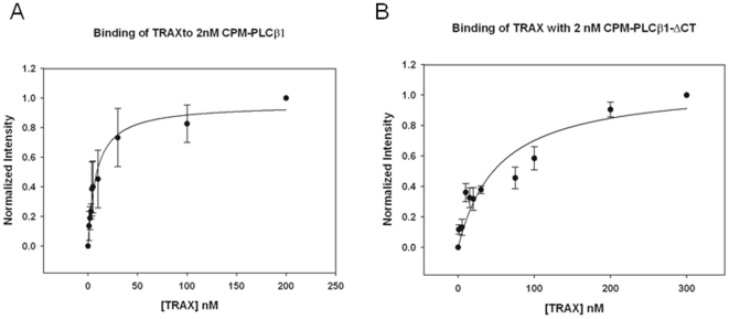
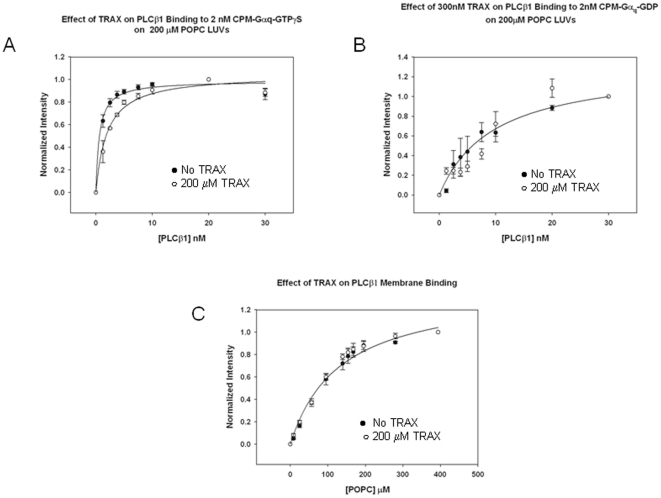
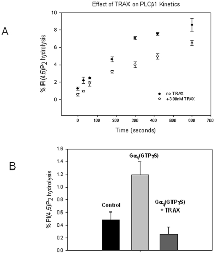
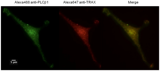
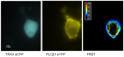
Mammalian phospholipase Cβ1 (PLCβ1) is activated by the ubiquitous Gα(q) family of G proteins on the surface of the inner leaflet of plasma membrane where it catalyzes the hydrolysis of phosphatidylinositol 4,5 bisphosphate. In general, PLCβ1 is mainly localized on the cytosolic plasma membrane surface, although a substantial fraction is also found in the cytosol and, under some conditions, in the nucleus. The factors that localize PLCβ1in these other compartments are unknown. Here, we identified a novel binding partner, translin-associated factor X (TRAX). TRAX is a cytosolic protein that can transit into the nucleus. In purified form, PLCβ1 binds strongly to TRAX with an affinity that is only ten-fold weaker than its affinity for its functional partner, Gα(q). In solution, TRAX has little effect on the membrane association or the catalytic activity of PLCβ1. However, TRAX directly competes with Gα(q) for PLCβ1 binding, and excess TRAX reverses Gα(q) activation of PLCβ1. In C6 glia cells, endogenous PLCβ1 and TRAX colocalize in the cytosol and the nucleus, but not on the plasma membrane where TRAX is absent. In Neuro2A cells expressing enhanced yellow and cyano fluorescent proteins (i.e., eYFP- PLCβ1 and eCFP-TRAX), Förster resonance energy transfer (FRET) is observed mostly in the cytosol and a small amount is seen in the nucleus. FRET does not occur at the plasma membrane where TRAX is not found. Our studies show that TRAX, localized in the cytosol and nucleus, competes with plasma-membrane bound Gα(q) for PLCβ1 binding thus stabilizing PLCβ1 in other cellular compartments.


Conflict of interest statement

Competing Interests: The authors have declared that no competing interests exist.

Figures

Figure 1
Figure 1. The structure of TRAX is mainly helical.
Circular dichroism spectrum of 20 µM TRAX in 20 mM Hepes, 160 mM NaCl, pH 7.4.
Figure 2
Figure 2. TRAX binds strongly to PLCβ1.
A – Binding of TRAX to 2 nM CPM- PLCβ1 as monitored by the increase in CPM intensity where the normalized fluorescence intensity is shown as a function of TRAX concentration. In these studies, an 80% increase in intensity was observed as compared to control samples that substituted buffer for TRAX. Also shown is the fitted curve to a bimolecular dissociation constant where Kd = 8±1 nM (n = 6 and S.D. is shown). B – Identical study as 2A except that the COOH-terminal deletion mutant of PLCβ1 (PLCβ1-ΔC) was used instead of the full length enzyme (n = 3 and S.D. is shown). While a binding curve is shown to guide the eye, the affinity between the proteins was too weak to be accurately fit to a bimolecular dissociation constant. We note that the total change in CPM intensity was also ∼80% at the end of the titration.
Figure 3
Figure 3. TRAX competes with Gαq for PLCβ1 binding but not membranes.
A – Binding of PLCβ1 to 2 nM activated CPM- Gαq(GTPγS) in the absence (•) and presence (○) of 200 nM TRAX right panel showing the loss in Gαq affinity when TRAX is present, where n = 6 and S.D. is shown. We note that an ∼20% increase in CPM intensity was seen both without and with TRAX. B- Binding of PLCβ1 to 2 nM deactivated CPM- Gαq(GDP) in the absence (•) and presence (○) of 200 nM TRAX, where n = 3 and S.D. is shown. C- Binding of PLCβ1 to PC∶PS∶PE (1∶1∶1) large, unilamellar vesicles in the absence (•) (Kp = 132 µM) and presence (○) (Kp = 120 µM) of 200 nM TRAX as measured by the increase in CPM intensity as LUVs are titrated into the solution, where n = 3 and S.D. is shown.
Figure 4
Figure 4. TRAX affects the activation of PLCβ1 by Gαq.
A – The effect of 300 nM TRAX on the rate of PI(4,5)P2 hydrolysis catalyzed by 25nM PLCβ1 (n = 3 and S.D. is shown). As can be seen, TRAX does not affect the initial velocity of the curve. B- Prevention of activation of 5 nM PLCβ1 by 5nM Gαq by 300 nM TRAX, where n = 8 and S.D. is shown.
Figure 5
Figure 5. TRAX and PLCβ1 co-localize in C6 glial cells.
Example of a co-immunofluorescence study of endogenous PLCβ1 (left panel) as visualized by Alexa488-labeled antibody, TRAX (middle panel) visualized by Alexa647-labeled antibody and the resulting merged image (right panel) in C6 glial cells. The scale bar is 5 µm.
Figure 6
Figure 6. TRAX associates with PLCβ1 in N2A cells.
Example of a FRET study showing the raw images of eYFP- PLCβ1 (left panel), eCFP-TRAX (middle panel) and the normalized FRET image (right panel) in transfected Neuro2A cells where amount of FRET is determined by the sensitized emission (see . The scale bar is 5 µm.

References

    1. Suh P, Park J, Manzoli L, Cocco L, Peak J, et al. Multiple roles of phosphoinositide-specific phospholipase C isozymes. BMB reports. 2008;41:415–434. - PubMed
    1. Rebecchi MJ, Pentyala SN. Structure, function, and control of phosphoinositide-specific phospholipase C. PhysiolRev. 2000;80:1291–1335. - PubMed
    1. Exton JH. Regulation of phosphoinositide phospholipases by hormones, neurotransmitters, and other agonists linked to G proteins. Annual Review of Pharmacology & Toxicology. 1996;36:481–509. - PubMed
    1. Rhee SG. Regulation of phosphoinositide-specific phospholipase C. AnnRevBiochem. 2001;70:281–312. - PMC - PubMed
    1. Runnels LW, Jenco J, Morris A, Scarlata S. Membrane binding of phospholipases C-b1 and C-b2 is independent of phosphatidylinositol 4,5-bisphosphate and the a and bg subunits of G proteins. Biochemistry. 1996;35:16824–16832. - PubMed

Publication types

MeSH terms

LinkOut - more resources