. 2010 Nov 29;5(11):e14110.
doi: 10.1371/journal.pone.0014110.

Estrogen receptor β exerts tumor repressive functions in human malignant pleural mesothelioma via EGFR inactivation and affects response to gefitinib

Affiliations

Estrogen receptor β exerts tumor repressive functions in human malignant pleural mesothelioma via EGFR inactivation and affects response to gefitinib

Giulia Pinton et al. PLoS One. .

Abstract

Background: The role of estrogen and estrogen receptors in oncogenesis has been investigated in various malignancies. Recently our group identified estrogen receptor beta (ERβ) expression as an independent prognostic factor in the progression of human Malignant Pleural Mesothelioma (MMe), but the underlying mechanism by which ERβ expression in tumors determines clinical outcome remains largely unknown. This study is aimed at investigating the molecular mechanisms of ERβ action in MMe cells and disclosing the potential translational implications of these results.

Methods: We modulated ERβ expression in REN and MSTO-211H MMe cell lines and evaluated cell proliferation and EGF receptor (EGFR) activation.

Results: Our data indicate that ERβ knockdown in ER positive cells confers a more invasive phenotype, increases anchorage independent proliferation and elevates the constitutive activation of EGFR-coupled signal transduction pathways. Conversely, re-expression of ERβ in ER negative cells confers a more epithelioid phenotype, decreases their capacity for anchorage independent growth and down-modulates proliferative signal transduction pathways. We identify a physical interaction between ERβ, EGFR and caveolin 1 that results in an altered internalization and in a selective reduced activation of EGFR-coupled signaling, when ERβ is over-expressed. We also demonstrate that differential expression of ERβ influences MMe tumor cell responsiveness to the therapeutic agent: Gefitinib.

Conclusions: This study describes a role for ERβ in the modulation of cell proliferation and EGFR activation and provides a rationale to facilitate the targeting of a subgroup of MMe patients who would benefit most from therapy with Gefitinib alone or in combination with Akt inhibitors.


Conflict of interest statement

Competing Interests: The authors have declared that no competing interests exist.

Figures

Figure 1
Figure 1. ERβ expression in ERs negative MMe cells reduces their growth rate.
A) Western Blot analysis of cell extracts from mock- and ERβ expressing MSTO-211H cells. Representative of three separate experiments. B) Upper panels show phase contrast microphotographs (200X magnification) of mock- or ERβ-transfected MSTO-211H cells, visualizing the acquisition of a more epithelioid phenotype in transfected cells. Lower panels show cells fixed in ethanol and stained for actin with phalloidin–TRITC as described. Note the actin rearrangement in ERβ expressing cells (400X magnification). C) Cell proliferation curves of mock- and ERβ-transfected MSTO-211H cells cultured in complete medium for 24 and 48 hours. Each value represents mean ± SD (n = 3). D) Total soft agar colony counts for mock- or ERβ-transfected MSTO-211H cells were done by three independent investigators microscopically visualizing individual colonies (clusters of 15 or more cells) in 10 random microscopic fields. Columns represent the fold increase of the mean number of colonies in 10 fields; bars, SD; * p<0.05. Representative of three separate experiments.
Figure 2
Figure 2. ERβ silencing promotes MMe cell proliferation.
A) Western Blot analysis of cell extracts from mock- and ERβ silenced REN cells. Representative of three separate experiments. B) Upper panels show phase contrast microphotographs (200X magnification) of mock- or shERβ-transfected REN cells, visualizing the loss of contact inhibition and formation of foci in vitro. Lower panels show cells fixed in ethanol and stained with phalloidin-TRITC to stain for actin as described. Note the actin rearrangement in ERβ silenced cells (400X magnification). C) Cell proliferation curves of mock- and shERβ-transfected REN cells cultured in complete medium for 24 and 48 hours. Each value represents mean ± SD (n = 3). D) Total soft agar colony counts for mock- or shERβ-transfected REN cells were done by three independent investigators microscopically visualizing individual colonies (clusters of 15 or more cells) in 10 random microscopic fields. Columns represent the fold increase of the mean number of colonies; bars, SD; * p<0.05. Representative of three separate experiments.
Figure 3
Figure 3. ERβ over-expression influences EGFR mediated signaling and internalization.
A) The graph show the growth curves of mock- and ERβ-transfected REN cell treated for 24 and 48 hours with 5 ng/ml of EGF in 2% FBS culture medium. At each time point, the cells were assayed for proliferation. Each value represents mean ± SD (n = 3). Adjacent to the graph is reported a representative Western blot analysis that documents ERβ expression. Tubulin staining indicates equal loading of the proteins. B) Mock- and ERβ- transfected REN cells made quiescent for 2 hours were treated with 5 ng/ml of EGF for 5 minutes and detergent extracted. Levels of phosphorylated EGFR, ERK 1/2 MAP kinases and Akt were analyzed by immunoblotting. Membranes were also blotted with antibodies to EGFR, Erk1/2 and Akt to evaluate protein expression. Tubulin was blotted to show equal amount of loading. Western blot analysis with anti ERβ antibodies documents its expression in transfected cells. Representative of three separate experiments. C) Evaluation of EGFR internalization was performed by Flow cytometry analysis on wild type and ERβ expressing REN cells treated 60 or 120 minutes with 10 ng/ml of human recombinant EGF. Histograms represent percentage of positive cells following incubation with anti-EGFR antibody indicated for each condition ± SD. Data are representative of three separate experiments. D) Representative immunoprecipitation experiment of membrane associated EGFR performed on mock and ERβ over-expressing REN cells, treated 60 or 120 minutes with 10 ng/ml of human recombinant EGF. Membrane was blotted with anti-pY and anti-EGFR antibodies.
Figure 4
Figure 4. ERβ associates with EGFR and caveolin 1.
A) Co-immunoprecipitation experiments were performed on REN cells treated 1 and 5 minutes with 5 ng/ml of human recombinant EGF. ERβ and caveolin 1 were detected by Western blot in immunoprecipitations of membrane associated EGFR. B) Co-immunoprecipitation experiments were performed on mock and ERβ over-expressing REN cells treated 5 minutes with 5 ng/ml of human recombinant EGF. ERβ and caveolin 1 were detected by Western blot in immunoprecipitations of membrane associated EGFR. C) Confocal double fluorescent microscopy analysis of red-labeled ERβ with green-labeled EGFR or caveolin 1 in mock- (left panel) or ERβ-transfected (right panel) REN cells treated or not 5 minutes with 5 ng/ml of human recombinant EGF. D) Confocal fluorescent microscopy analysis showing the localization of green-labeled EGFR and phalloidin-TRITC labeled actin filaments in mock and in ERβ and ERα transfected REN cells. Nuclei were counterstained with DAPI.
Figure 5
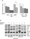
Figure 5. ERβ expression influences response of MMe cells to Gefitinib.
A) Effects of Gefitinib on viable number were evaluated in mock-, ERβ- and shERβ-transfected REN and in mock- and ERβ- transfected MSTO-211H cell lines. Cells were incubated in serum-containing medium in the presence of 5 µM Gefitinib for 24–48 hours. As control 0.1% DMSO vehicle alone was used. Results are expressed as number of viable cells relative to control at 48 hours of treatment; bars, ± SD; * p<0.05. Data are representative of three separate experiments. B) shERβ-transfected REN and MSTO-211H cells were treated with 5 ng/ml of EGF for 5 minutes in the absence or presence of 5 µM Gefitinib and detergent extracted. Levels of phosphorylated EGFR, ERK 1/2 MAP kinases and Akt were analyzed by immunoblot. Membranes were also blotted with antibodies to EGFR, Erk1/2, and Akt to evaluate protein expression. Tubulin was blotted to show equal amount of loading. Western blot analysis with anti ERβ antibodies documents expression in transfected cells. Representative of three separate experiments.

References

    1. Yang H, Testa JR, Carbone M. Mesothelioma epidemiology, carcinogenesis, and pathogenesis. Curr Treat Options Oncol. 2008;9:147–57. Review. - PMC - PubMed
    1. Pass HI, Carbone M. Current status of screening for malignant pleural mesothelioma. Semin Thorac Cardiovasc Surg. 2009;21:97–104. - PubMed
    1. Richards WG. Recent advances in mesothelioma staging. Semin Thorac Cardiovasc Surg. 2009;21:105–10. - PubMed
    1. Steele JP. Prognostic factors for mesothelioma. Hematol Oncol Clin North Am. 2005;19:1041–52. Review. - PubMed
    1. Pinton G, Brunelli E, Murer B, Puntoni R, Puntoni M, et al. Oestrogen receptor β impacts on prognosis of human malignant mesothelioma. Cancer Res. 2009;69:4598–604. - PubMed

Publication types

MeSH terms