. 2010 Nov 29;5(11):e15040.
doi: 10.1371/journal.pone.0015040.

Freshwater sponges have functional, sealing epithelia with high transepithelial resistance and negative transepithelial potential

Affiliations

Freshwater sponges have functional, sealing epithelia with high transepithelial resistance and negative transepithelial potential

Emily D M Adams et al. PLoS One. .

Abstract

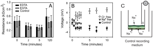
Epithelial tissue - the sealed and polarized layer of cells that regulates transport of ions and solutes between the environment and the internal milieu - is a defining characteristic of the Eumetazoa. Sponges, the most ancient metazoan phylum, are generally believed to lack true epithelia, but their ability to occlude passage of ions has never been tested. Here we show that freshwater sponges (Demospongiae, Haplosclerida) have functional epithelia with high transepithelial electrical resistance (TER), a transepithelial potential (TEP), and low permeability to small-molecule diffusion. Curiously, the Amphimedon queenslandica sponge genome lacks the classical occluding genes [5] considered necessary to regulate sealing and control of ion transport. The fact that freshwater sponge epithelia can seal suggests that either occluding molecules have been lost in some sponge lineages, or demosponges use novel molecular complexes for epithelial occlusion; if the latter, it raises the possibility that mechanisms for occlusion used by sponges may exist in other metazoa. Importantly, our results imply that functional epithelia evolved either several times, or once, in the ancestor of the Metazoa.


Conflict of interest statement

Competing Interests: The authors have declared that no competing interests exist.

Figures

Figure 1
Figure 1. Structure of the sponge epithelium.
(A) Diagram of the experimental chamber, and (B) scanning electron microscopy (SEM) of sponge tissue showing: the bilayered dermal tissues (dt), suspended over the subdermal space (ss) by shafts of spicules (s); the choanosome (ch) with the choanocyte pumps; excurrent canals (ec); and an osculum (o). For transepithelial recordings, apical (AP) and basolateral (BL) compartments were effectively separated by two confluent cell layers - the basopinacoderm (bp) and endopinacoderm (enp) - surrounding a thin mesohyl (me) (inset); scale bar b, 100 µm. (C) SEM of the dermal tissue shows exopinacocytes are very close to one-another (opposing arrows show cell-cell spacing); scale 1 µm. (D) Fluorescent labelling of the basopinacoderm with the steryl dye FM 1–43 highlights the borders of cells showing that a confluent layer of cells covered culture membranes; scale 10 µm. (E) Transmission electron microscopy of exopinacocytes shows tight membrane apposition between adjacent cells (arrow). These areas were associated with 10 nm diameter cytoskeletal fibres (arrowheads); scale 200 nm. (F) Fluorescent labelling using phallacidin reveals dense plaques of actin (arrowhead) between cells; nuclei, blue; scale, 5 µm.
Figure 2
Figure 2. Electrical resistance and impermeability of S. lacustris epithelia.
(A) The resistance to ion passage of Spongilla epithelia derived from aggregates (Ag) (1098.5 Ω cm2±671.5 s.e. n = 33) and gemmules (Gm) (1932.5 Ω cm2±253.5 s.e. n = 22) was comparable to the resistance of epithelia from vertebrate tissues (MPT =  mammal proximal tubule, MGB =  mammal gall bladder, SPT =  salamander proximal tubule, RC =  rabbit colon, MDT  =  mammal kidney distal tubule FTS =  frog/toad skin; FWG =  freshwater fish gill; TUB =  toad urinary bladder). (B) Permeability to 3H inulin decreased with increasing resistance of sponge cultures. (C) 3H inulin gradually accumulated on the basolateral side of cultures with low resistance (solid circles, <500 Ω cm2), but was excluded by high resistance epithelia (open circles, >800 Ω cm2). (D) Regions of membrane fusion, as seen by transmission electron microscopy (TEM) were common near the most apical point of cell contact in exopinacocytes; scale 100 nm. (E) Ruthenium red (RR, the dark precipitate) was excluded from paracellular clefts (double arrow); scale, 200 nm. (F) RR was excluded by exopinacocytes (ExP) and endopinacocytes (EnP) from the collagenous (Co) internal mesohyl containing mesohyl cells (M). The tracer entered the aquiferous system though ostia and therefore coated the endopinacoderm (En) roof of the subdermal space (SS); scale, 500 nm.
Figure 3
Figure 3. Ionic basis of occlusion in sponge epithelia.
(A) Effect of EGTA or EDTA treatment on resistance. All cultures lost resistance in the recording medium and recovered high resistance after 2 hr in the culture-medium. (B) Voltage across the epithelium was negative in the normal culture medium and in sodium-free medium and positive in chloride-free medium. (C) Schematic showing the uptake of sodium in normal recording medium to generate a negative potential.
Figure 4
Figure 4. Sponge phylogeny and the evolution of epithelial characters.
A phylogeny of Metazoa adapted from Philippe et al 2009 which considers Porifera monophyletic. Sponge phylogeny is adapted from . Freshwater sponges belong to the group indicated with a box (G4). Letters indicate evidence of epithelial characters: (a) transepithelial resistance (TER) and transepithelial potential (TEP) in freshwater sponges; (b) septate junctions in calcareous sponges; (c) a basement membrane in homoscleromorphs; (d) absence (loss?) of a basement membrane in placozoans; (e) true epithelia with septate junctions, a basal lamina, TER and TEP in cnidarians and bilaterians. Arrows indicate three potential origins of epithelia; the solid bar indicates the most parsimonious scenario for the origin of epithelia.

References

    1. Philippe H, Derelle R, Lopez P, Pick K, Borchiellini C, et al. Phylogenomics Revives Traditional Views on Deep Animal Relationships. Curr Biol. 2009;19:706–712. - PubMed
    1. Love GD, Grosjean E, Stalvies C, Fike DA, Grotzinger JP, et al. Fossil steroids record the appearance of Demospongiae during the Cryogenian period. Nature. 2009;457:718–721. - PubMed
    1. Mackie GO. Introduction to the diploblastic level. In: Bereiter-Hahn J, Matoltsy AG, Richards KS, editors. Biology of the integument. Berlin: Springer-Verlag; 1984. pp. 43–46.
    1. Cereijido M, Contreras RG, Shoshani L. Cell adhesion, polarity, and epithelia in the dawn of metazoans. Physiol Rev. 2004;84:1229–1262. - PubMed
    1. Srivastava M, Simakov O, Chapman J, Fahey B, Gauthier M, et al. The Amphimedon queenslandica genome and the evolution of animal complexity. Nature. 2010;466:720–727. - PMC - PubMed

Publication types