. 2010 Nov 24;5(11):e14108.
doi: 10.1371/journal.pone.0014108.

Overrepresentation of IL-17A and IL-22 producing CD8 T cells in lesional skin suggests their involvement in the pathogenesis of psoriasis

Affiliations

Overrepresentation of IL-17A and IL-22 producing CD8 T cells in lesional skin suggests their involvement in the pathogenesis of psoriasis

Pieter C M Res et al. PLoS One. .

Abstract

Background: Although recent studies indicate a crucial role for IL-17A and IL-22 producing T cells in the pathogenesis of psoriasis, limited information is available on their frequency and heterogeneity and their distribution in skin in situ.

Methodology/principal findings: By spectral imaging analysis of double-stained skin sections we demonstrated that IL-17 was mainly expressed by mast cells and neutrophils and IL-22 by macrophages and dendritic cells. Only an occasional IL-17(pos), but no IL-22(pos) T cell could be detected in psoriatic skin, whereas neither of these cytokines was expressed by T cells in normal skin. However, examination of in vitro-activated T cells by flow cytometry revealed that substantial percentages of skin-derived CD4 and CD8 T cells were able to produce IL-17A alone or together with IL-22 (i.e. Th17 and Tc17, respectively) or to produce IL-22 in absence of IL-17A and IFN-γ (i.e. Th22 and Tc22, respectively). Remarkably, a significant proportional rise in Tc17 and Tc22 cells, but not in Th17 and Th22 cells, was found in T cells isolated from psoriatic versus normal skin. Interestingly, we found IL-22 single-producers in many skin-derived IL-17A(pos) CD4 and CD8 T cell clones, suggesting that in vivo IL-22 single-producers may arise from IL-17A(pos) T cells as well.

Conclusions/significance: The increased presence of Tc17 and Tc22 cells in lesional psoriatic skin suggests that these types of CD8 T cells play a significant role in the pathogenesis of psoriasis. As part of the skin-derived IL-17A(pos) CD4 and CD8 T clones developed into IL-22 single-producers, this demonstrates plasticity in their cytokine production profile and suggests a developmental relationship between Th17 and Th22 cells and between Tc17 and Tc22 cells.


Conflict of interest statement

Competing Interests: The authors have declared that no competing interests exist.

Figures

Figure 1
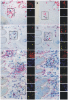
Figure 1. IL-17 and IL-22 expression in psoriatic skin.
Microscopic detail of lesional psoriatic skin stained for IL-17 (A–D) or IL-22 (E–H) in combination with CD3 (A, B, E), myeloperoxidase (C), tryptase (D, H), CD11c (F), or CD68 (G). Cytokines are visualized in blue and all different cell markers in red. Each set of three small pictures on the right side represents a composite fluorescent-like image in pseudo-colors of the rectangle-marked area and was obtained after unmixing the individual colors red and blue with spectral imaging. Colocalization of red and blue is indicated in yellow. Spectral imaging analysis of this double-stained series of sections clearly shows the presence of an occasional IL-17pos T cell in psoriatic lesions, whereas IL-22pos T cells were absent. The IL-17 expression in psoriatic skin was predominantly confined to neutrophils and mast cells and IL-22 was highly expressed in many dendritic cells and in most macrophages. The figure is representative of the results from three different donors. Examples of double-stained cells are indicated with an arrow and single-stained cells with an arrowhead. Original magnification 200×.
Figure 2
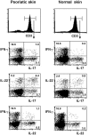
Figure 2. Cytokine expression in skin derived dermal T cells after in vitro stimulation.
T cells with the ability to produce IL-17A and/or IL-22 and/or IFN-γ are present in the dermis of both psoriatic and normal skin. Bulk T cells derived from the dermis were stimulated with PMA and ionomycin and stained for cell surface expression of CD3, CD4 and CD8 and intracellular expression of IL-17A, IL-22 and IFN-γ. Dot-plots contain the CD3 cells within the lymphocyte gate. This analysis shows the presence among dermal CD3 T cells of both normal and psoriatic skin of single and double producers of IL-17A, IL-22 and IFN-γ. Results are representative for eight independent experiments.
Figure 3
Figure 3. Cytokine profiles of dermis-derived CD4 and CD8 bulk T cell populations.
Relative contribution of T cells with different expression profiles of IL-17A, IL-22 and IFN-γ production among in vitro activated dermal CD4 T cells (a) and CD8 T (b) from psoriatic skin (closed symbols) versus normal skin (open symbols). Each series of a particular symbol represents data from one individual. Specifically T cells with the ability to produce IL-17A and/or IL-22 are present in increased percentages of the CD8 T cell population in lesional skin compared to normal skin. *P<0.05; **P<0.01 Depicted data show the results obtained by six color flow cytometry of the eight independent experiments.
Figure 4
Figure 4. Proportions of IL-17A and IL-22 producing T cells in the epidermis versus dermis.
The increase in the percentages of Tc17 and IL-22 producing CD8 T cells is even more pronounced in the psoriatic epidermis than in the psoriatic dermis, whereas normal epidermis and dermis are almost devoid of such T cells (n = 4).
Figure 5
Figure 5. Cytokine profile of psoriatic skin derived Th17 and Tc17 clones.
IL-17Apos CD4 and CD8 T cell clones (Th17 and Tc17, respectively) from psoriatic skin have the ability to give rise to a proportion of IL-22 producing cells that lack IL-17A and IFN-γ expression (the putative Th22 and Tc22, respectively). IL-17Apos CD4 dermal T cells and CD8 epidermal T cells derived from psoriatic skin were cloned and subsequently assayed for intracellular IL-17A, IL-22, and IFN-γ after PMA ionomycin stimulation. Representative examples of CD4 and CD8 T cell clones are given. Part of the cells within the CD4 Th17 clone expressed IL-22, but lacked both IL-17A (right-bottom quadrant in the upper dot-plot) and IFN-γ expression (associated histogram indicated by arrow), a cytokine pattern typical of Th22 cells. Likewise, part of the cells of the CD8 Tc17 clone (bottom dot-plot) lacked IL-17A and IFN-γ expression, which is a Tc22 cytokine profile.

References

    1. Teunissen MB, Piskin G, Res PC, de Groot M, Picavet DI, et al. State of the art in the immunopathogenesis of psoriasis. G Ital Dermatol Venereol. 2007;142:229–242.
    1. Bos JD, de Rie MA, Teunissen MB, Piskin G. Psoriasis: dysregulation of innate immunity. Br J Dermatol. 2005;152:1098–1107. - PubMed
    1. Bos JD. Psoriasis, innate immunity, and gene pools. J Am Acad Dermatol. 2007;56:468–471. - PubMed
    1. Nickoloff BJ, Xin H, Nestle FO, Qin JZ. The cytokine and chemokine network in psoriasis. Clin Dermatol. 2007;25:568–573. - PubMed
    1. Austin LM, Ozawa M, Kikuchi T, Walters IB, Krueger JG. The majority of epidermal T cells in Psoriasis vulgaris lesions can produce type 1 cytokines, interferon-gamma, interleukin-2, and tumor necrosis factor-alpha, defining TC1 (cytotoxic T lymphocyte) and TH1 effector populations: a type 1 differentiation bias is also measured in circulating blood T cells in psoriatic patients. J Invest Dermatol. 1999;113:752–759. - PubMed

MeSH terms