. 2010 Nov 24;5(11):e14109.
doi: 10.1371/journal.pone.0014109.

Suppressive effects of vascular endothelial growth factor-B on tumor growth in a mouse model of pancreatic neuroendocrine tumorigenesis

Affiliations

Suppressive effects of vascular endothelial growth factor-B on tumor growth in a mouse model of pancreatic neuroendocrine tumorigenesis

Imke Albrecht et al. PLoS One. .

Abstract

Background: The family of vascular endothelial growth factors (VEGF) contains key regulators of blood and lymph vessel development, including VEGF-A, -B, -C, -D, and placental growth factor. The role of VEGF-B during physiological or pathological angiogenesis has not yet been conclusively delineated. Herein, we investigate the function of VEGF-B by the generation of mouse models of cancer with transgenic expression of VEGF-B or homozygous deletion of Vegfb.

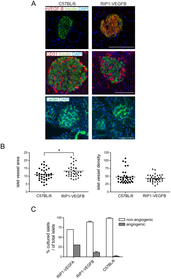
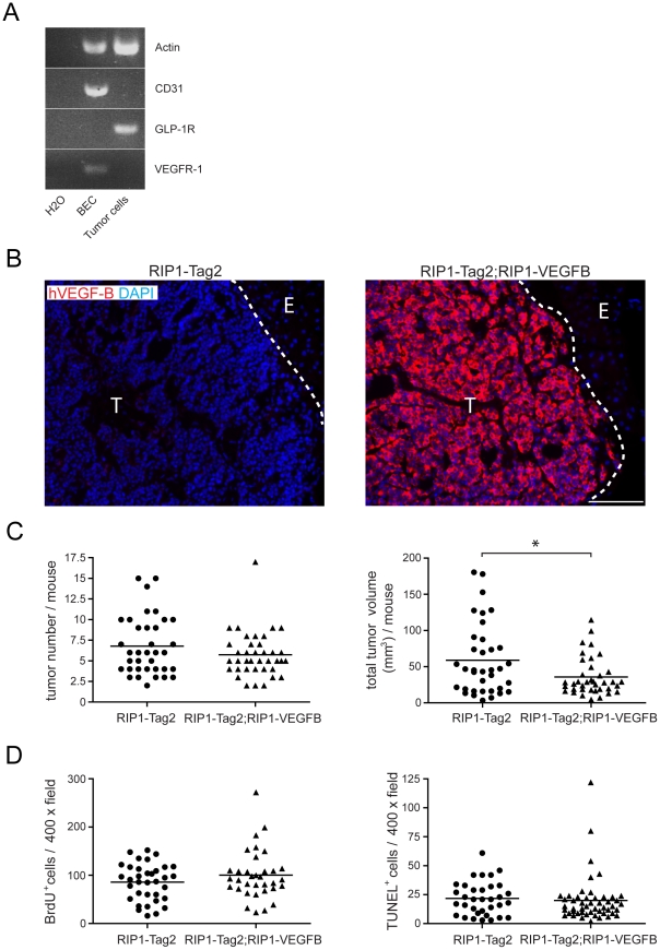
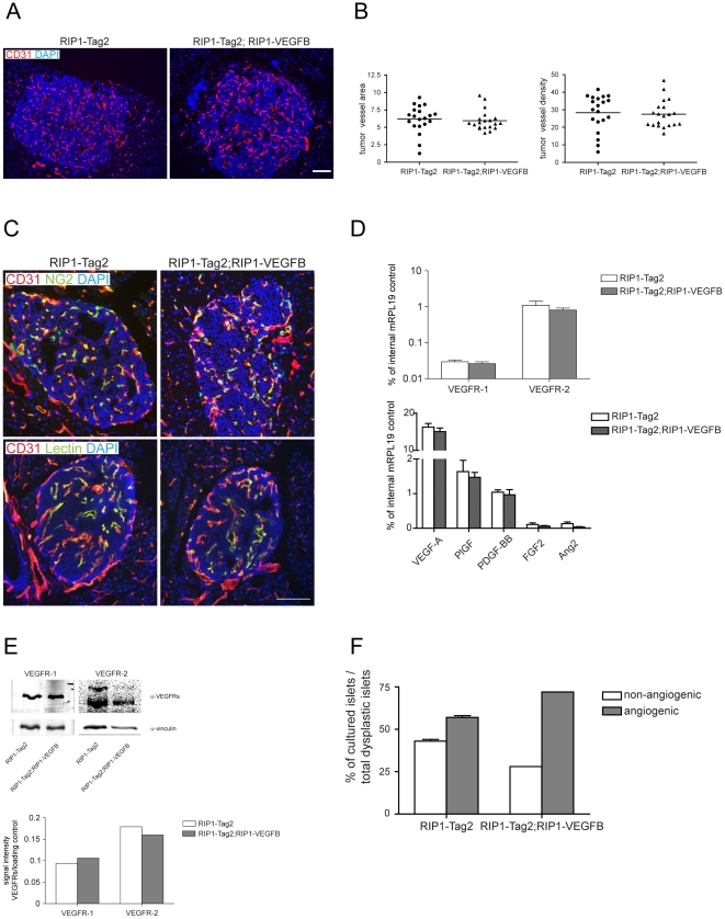
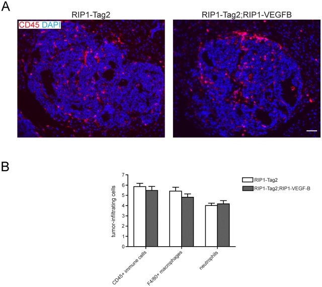
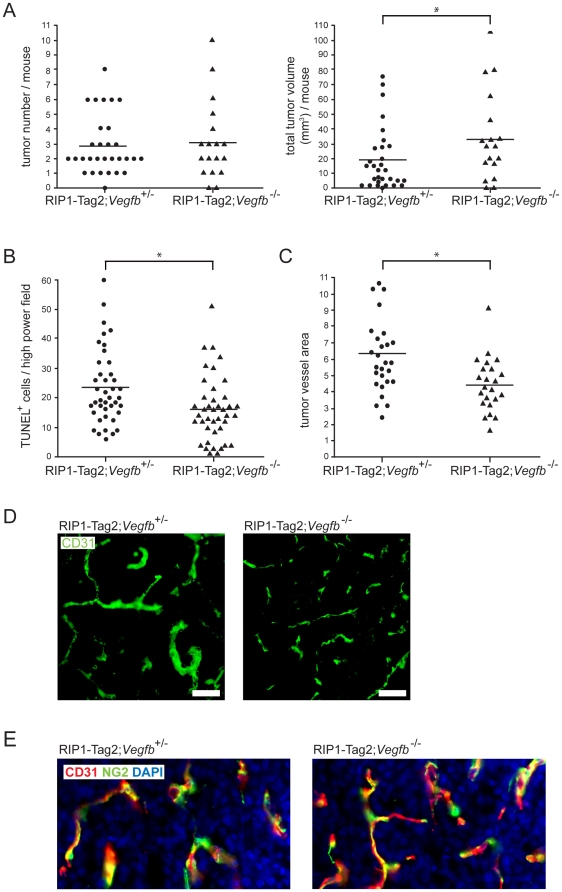
Methodology/principal findings: Ectopic expression of VEGF-B in the insulin-producing β-cells of the pancreas did not alter the abundance or architecture of the islets of Langerhans. The vasculature from transgenic mice exhibited a dilated morphology, but was of similar density as that of wildtype mice. Unexpectedly, we found that transgenic expression of VEGF-B in the RIP1-Tag2 mouse model of pancreatic neuroendocrine tumorigenesis retarded tumor growth. Conversely, RIP1-Tag2 mice deficient for Vegfb presented with larger tumors. No differences in vascular density, perfusion or immune cell infiltration upon altered Vegfb gene dosage were noted. However, VEGF-B acted to increase blood vessel diameter both in normal pancreatic islets and in RIP1-Tag2 tumors.

Conclusions/significance: Taken together, our results illustrate the differences in biological function between members of the VEGF family, and highlight the necessity of in-depth functional studies of VEGF-B to fully understand the effects of VEGFR-1 inhibitors currently used in the clinic.


Conflict of interest statement

Competing Interests: The authors have declared that no competing interests exist.

Figures

Figure 1
Figure 1. Characterization of angiogenesis in pancreatic islets from RIP1-VEGFB mice.
A) Pancreatic sections of control C57BL/6 (left) and of RIP1-VEGFB mice (right) were stained for human VEGF-B (red) to detect transgene expression (upper panel), for CD31 (red) to examine intra-insular blood vessel distribution (middle panel) and were perfusion stained with FITC-coupled tomato lectin to evaluate intra-insular blood vessel functionality (lower panel). To visualize islets of Langerhans, pancreatic sections were co-stained with insulin. Nuclei were visualized by DAPI stain. Scale bar: 100 µm. B) Quantification of islet microvessel area and density of C57BL/6 (N =  5, n =  37) and RIP1-VEGFB (N =  4, n =  36) mice. Analysis was performed by determination of the CD31 stained area (left panel) or CD31 counts (right panel) in relation to the islet area using computer-assisted image analysis. * P =  0.0112. N =  number of analyzed mice, n =  number of islets. C) Islets isolated from RIP1-VEGF-A (n = 23, N = 2), RIP1-VEGFB167 (n = 60, N = 10) and C57BL/6 (n = 38, N = 9), mice were co-cultured with HUVEC in a collagen gel matrix and their ability to induce an angiogenic response was determined. The data points represent the average from two independent experiments using C57Bl/6 and RIP1-VEGFB167 mice, while all islets from RIP1-VEGFA mice were analyzed in a single experiment. n =  number of islets, N =  number of mice.
Figure 2
Figure 2. Characterization of the phenotype of tumors from RIP1-Tag2; RIP1-VEGFB mice.
A) RT-PCR analysis of VEGF-R1 expression in GLP1R+ β-tumor-cells and CD31+ tumor-derived blood-endothelial cells (BEC) isolated from 12 weeks old RIP1-Tag2 mice. B) Pancreatic tumor sections of control RIP1-Tag2 (left) and RIP1-Tag2; RIP1-VEGFB (right) mice were stained for human VEGF-B (red) to detect transgene expression. Nuclei were counterstained with DAPI. T =  Tumor, E =  Exocrine pancreas. Scale bar: 100 µm. C) Tumor incidence (left) and volumes (right) of RIP1-Tag2 (N =  36) and RIP1-Tag2; RIP1-VEGFB (N =  38) mice were determined at the age of 12 weeks. Single points represent the total tumor volume (or tumor number) per mouse as indicated. * P  =  0.0149 (Student's t-test). D) Tumor cell proliferation (left) and apoptosis (right) in RIP1-Tag2 and RIP1-Tag2; RIP1-VEGFB mice was determined by counting the number of BrdU and TUNEL positive tumor cells in a total of 7 to 10 microscopic fields (magnification 400×) per mouse.
Figure 3
Figure 3. Characterization of the vascular and angiogenic profile of tumors derived from RIP1-Tag2; RIP1-VEGFB mice.
A) Representative immunofluorescence microphotographs of pancreatic tumor sections of RIP1-Tag2 (left) and RIP1-Tag2; RIP1-VEGFB (right) mice stained for CD31. Scale bar: 100 µm. B) Quantification of intratumoral vessel density in RIP1-Tag2 (N =  5, n =  20) and RIP1-Tag2; RIP1-VEGFB (N =  5, n =  20) was performed using computer-assisted image analysis. Results are displayed as relation of CD31 stained area or CD31 positive cell counts to tumor area. N =  number of analyzed mice, n =  number of islets. C) Representative immunofluorescence microphotographs of pancreatic tumor sections of RIP1-Tag2 (left) and of RIP1-Tag2; RIP1-VEGFB (right) mice double-stained with CD31 and the pericyte marker NG2 (upper panel) or with FITC-coupled tomato lectin (lower panel). Scale bar: 100 µm. D) Evaluation of mVEGFR-1, mVEGFR-2, mVEGF-A, mPlGF, mPDGF-BB, mFGF2 and mAng2 mRNA expression by quantitative RT-PCR in total tumors of RIP1-Tag2 (n = 5) and RIP1-Tag2; RIP1-VEGFB (n = 5) mice. The mRNA expression profiles of the indicated genes are normalized to the expression of the internal control gene ribosomal protein 19 (mRPL19). E) Upper panel: Immunoblot for mVEGFR-1, mVEGFR-2 and vinculin (loading control) of total tumor lysate prepared from pancreatic tumors of a 12 weeks-old RIP1-Tag2 and RIP1-Tag2; RIP1-VEGFB. Lower panel: Quantitation. F) Determination of the percentage of dysplastic islets isolated from RIP1-Tag2 (white bar, n =  24) and RIP1-Tag2; RIP1-VEGFB mice (grey bar, n =  25) which are able to induce an angiogenic response when they were co-cultured with HUVECs in a collagen gel matrix.
Figure 4
Figure 4. Analysis of inflammatory cell infiltration in tumors derived from RIP1-Tag2; RIP1-VEGFB mice.
A) Representative immunofluorescence microphotographs of pancreatic tumor sections of RIP1-Tag2 (left) and RIP1-Tag2; RIP1-VEGFB (right) mice stained for CD45. Scale bar: 100 µm. B) Quantification of tumor-infiltrating immune cells in RIP1-Tag2 (white bar, N =  5, n =  26-46) and RIP1-Tag2; RIP1-VEGFB (grey bar, N =  5, n = 27–45). The number of tumor-infiltrating CD45+ and F4/80+ cells as well as 7/4+ neutrophils was determined by quantification of the area stained for the selected marker in relation to the total tumor area using computer-assisted image analysis. N =  number of analyzed mice, n =  number of tumors.
Figure 5
Figure 5. Characterization of the tumor and angiogenic phenotype of tumors derived from Vegfb-deficient RIP1-Tag2 mice.
A) Quantification of tumor number (left) and total tumor burden (right) of 12 weeks-old RIP1-Tag2; Vegfb +/− (n = 30) and RIP1-Tag2; Vegfb −/− (n = 18) mice. B) Quantification of tumor cell apoptosis in lesions (n = 40 for each genotype) from RIP1-Tag2; Vegfb +/− and RIP1-Tag2; Vegfb −/− mice, as assessed by TUNEL assay. C,D) Vascular density (C) and morphology (D) in lesions from RIP1-Tag2; Vegfb +/− (n = 27) and RIP1-Tag2; Vegfb −/− (n = 23) mice. Scale bar, 50 µm. E) Pericyte coverage (n = 24 for each genotype), as visualized by immunostaining for the pericyte marker NG2 (green) in relation to the endothelial cell marker CD31 (red). Cell nuclei were visualized using DAPI (blue).

References

    1. Carmeliet P. Angiogenesis in life, disease and medicine. Nature. 2005;438:932–936. - PubMed
    1. Olsson AK, Dimberg A, Kreuger J, Claesson-Welsh L. VEGF receptor signalling - in control of vascular function. Nat Rev Mol Cell Biol. 2006;7:359–371. - PubMed
    1. Carmeliet P, Ferreira V, Breier G, Pollefeyt S, Kieckens L, et al. Abnormal blood vessel development and lethality in embryos lacking a single VEGF allele. Nature. 1996;380:435–439. - PubMed
    1. Ferrara N, Carver-Moore K, Chen H, Dowd M, Lu L, et al. Heterozygous embryonic lethality induced by targeted inactivation of the VEGF gene. Nature. 1996;380:439–442. - PubMed
    1. Fischer C, Jonckx B, Mazzone M, Zacchigna S, Loges S, et al. Anti-PlGF inhibits growth of VEGF(R)-inhibitor-resistant tumors without affecting healthy vessels. Cell. 2007;131:463–475. - PubMed

Publication types

MeSH terms

Substances