. 2010 Nov 23;5(11):e15535.
doi: 10.1371/journal.pone.0015535.

Quantitative high-throughput screening identifies 8-hydroxyquinolines as cell-active histone demethylase inhibitors

Affiliations

Quantitative high-throughput screening identifies 8-hydroxyquinolines as cell-active histone demethylase inhibitors

Oliver N F King et al. PLoS One. .

Abstract

Background: Small molecule modulators of epigenetic processes are currently sought as basic probes for biochemical mechanisms, and as starting points for development of therapeutic agents. N(ε)-Methylation of lysine residues on histone tails is one of a number of post-translational modifications that together enable transcriptional regulation. Histone lysine demethylases antagonize the action of histone methyltransferases in a site- and methylation state-specific manner. N(ε)-Methyllysine demethylases that use 2-oxoglutarate as co-factor are associated with diverse human diseases, including cancer, inflammation and X-linked mental retardation; they are proposed as targets for the therapeutic modulation of transcription. There are few reports on the identification of templates that are amenable to development as potent inhibitors in vivo and large diverse collections have yet to be exploited for the discovery of demethylase inhibitors.

Principal findings: High-throughput screening of a ∼236,000-member collection of diverse molecules arrayed as dilution series was used to identify inhibitors of the JMJD2 (KDM4) family of 2-oxoglutarate-dependent histone demethylases. Initial screening hits were prioritized by a combination of cheminformatics, counterscreening using a coupled assay enzyme, and orthogonal confirmatory detection of inhibition by mass spectrometric assays. Follow-up studies were carried out on one of the series identified, 8-hydroxyquinolines, which were shown by crystallographic analyses to inhibit by binding to the active site Fe(II) and to modulate demethylation at the H3K9 locus in a cell-based assay.

Conclusions: These studies demonstrate that diverse compound screening can yield novel inhibitors of 2OG dependent histone demethylases and provide starting points for the development of potent and selective agents to interrogate epigenetic regulation.


Conflict of interest statement

Competing Interests: CJS is a cofounder of ReOx, a company that is interested in the exploitation of the hypoxic response for therapeutic benefit. The authors declare no other competing interests. This does not alter the authors' adherence to all the PLoS ONE policies on sharing data and materials.

Figures

Figure 1
Figure 1. Screening and Hit Prioritization Strategy.
Figure 2
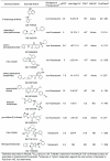
Figure 2. Representative HTS hits.
Shown are representative members of clusters along with results from the screen and prioritization experiments.
Figure 3
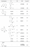
Figure 3. Structure-activity relationships for substituted 8-hydroxyquinolines identified in the qHTS against JMJD2E (qHTS IC50 values derived from the FDH-coupled assay).
Some substitution patterns were chosen for further medicinal chemistry; IC50s for these synthesized compounds are indicated in italics.
Figure 4
Figure 4. JMJD2A crystal structure complexed with 5-carboxy-8-HQ (SID 85736331).
(A) active site residues are in green sticks, 5-carboxy-8-hydroxyquinoline as cyan sticks, secondary structure helices in red and sheets in yellow. The Ni(II) ion (replacing Fe(II)), is a blue sphere and the Zn(II) is a grey sphere. (B) Binding mode of 5-carboxy-8-hydroxyquinoline in JMJD2A. The experimental 2Fo-Fc electron density is shown as grey mesh (contoured at 1σ). Hydrogen bonds to residues are shown in black. Hydrogen bonds involving waters (red spheres) are shown in red, distances in Å. (C) Overlay of JMJD2A crystal structure with the 5-carboxy-8-hydroxyquinoline (residues green, compound cyan, Ni(II) in blue) with the crystal structure with 2,4 PDCA (residues compound and Ni(II) ion are pink, PDB ID: 2VD7). The distances shown are for the 8-hydroxyquinoline structure between: 5-carboxy-8-hydroxyquinoline and His 276, Ni2+ and His 188 (in black), His 276 and Ni2+ (in red). Distances in Å. (D) Surface view of superimposition of 8-hydroxyquinoline 5-acid bound JMJD2A structure with that of JMJD2A bound to histone 3 lysine 9 trimethylated (H3K9me3) substrate peptide (grey and magenta sticks) and the 2-oxoglutarate (2OG) mimetic N-oxalylglycine (NOG, yellow sticks), PDB ID: 2OQ6. Binding of the 8-hydroxyquinoline 5-acid (cyan sticks) is likely to prevent binding of 2OG but not directly that of the H3K9me3 residue (in magenta) or other residues in the substrate peptide (in grey). Peptide sequence ARK(me3)STGGK(Ac). Ni2+ of structure 2OQ6, yellow sphere; Ni2+ of 8-hydroxyquinoline structure, green sphere. The triad of metal coordinating residues are in green sticks. Crystallographic data and refinement parameters are described in Table S2.
Figure 5
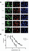
Figure 5. 5-Carboxy-8-HQ (SID 85736331) increases H3K9me3 levels in HeLa cells through inhibition of JMJD2A.
(A) Indirect immunofluorescence with anti-Flag (green), anti-H3K9me3 (red), and DAPI staining (blue) in HeLa cells overexpressing Flag-tagged JMJD2A. DMSO solvent treatment has no effect on JMJD2A demethylase activity (white arrows) while increasing concentrations of 5-carboxy-8-hydroxyquinoline treatment (100 µM to 300µM range shown) resulted in gradual increases in H3K9Me3 levels. The JMJD2A H188A enzymatic mutant does not affect H3K9Me3 levels when overexpressed. (B) Quantitation of H3K9me3 levels is shown as squares (▪). Standard deviations are derived from biological triplicates at inhibitor concentrations of 20 µM, 50 µM, 100 µM, 200 µM, 300 µM, and 400 µM treatments. Cytotoxicity (•) was assayed at 50 µM, 100 µM, 200 µM, 300 µM, 400 µM, 500 µM, 750 µM, 1 mM, 1,25 mM, 1.5 mM, 1.75 mM, 2 mM, 2.5 mM, and 3 mM.
Figure 6
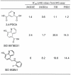
Figure 6. Inhibitor screening against JMJD2 and other human 2OG oxygenases.
IC50 values from MALDI-TOF MS assay. See Figure S4 for representative concentration-response curves and MALDI-TOF mass spectra.

References

    1. Ng S, Yue W, Oppermann U, Klose R. Dynamic protein methylation in chromatin biology. Cellular and Molecular Life Sciences. 2009;66:407–422. - PMC - PubMed
    1. Klose RJ, Zhang Y. Regulation of histone methylation by demethylimination and demethylation. Nat Rev Mol Cell Biol. 2007;8:307–318. - PubMed
    1. Mosammaparast N, Shi Y. Reversal of histone methylation: biochemical and molecular mechanisms of histone demethylases. Annu Rev Biochem. 2010;79:155–179. - PubMed
    1. Ruthenburg AJ, Allis CD, Wysocka J. Methylation of Lysine 4 on Histone H3: Intricacy of Writing and Reading a Single Epigenetic Mark. Molecular Cell. 2007;25:15–30. - PubMed
    1. Grewal SI, Jia S. Heterochromatin revisited. Nat Rev Genet. 2007;8:35–46. - PubMed

Publication types

MeSH terms