. 2010 Nov 23;5(11):e15447.
doi: 10.1371/journal.pone.0015447.

Evolution of allosteric citrate binding sites on 6-phosphofructo-1-kinase

Affiliations

Evolution of allosteric citrate binding sites on 6-phosphofructo-1-kinase

Aleksandra Usenik et al. PLoS One. .

Abstract

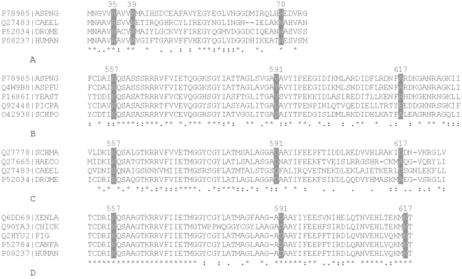
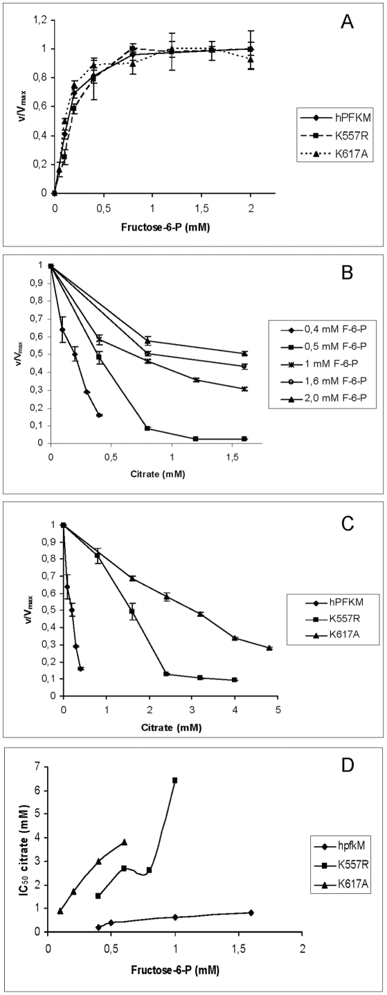
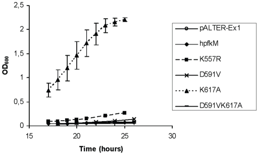
As an important part of metabolism, metabolic flux through the glycolytic pathway is tightly regulated. The most complex control is exerted on 6-phosphofructo-1-kinase (PFK1) level; this control overrules the regulatory role of other allosteric enzymes. Among other effectors, citrate has been reported to play a vital role in the suppression of this enzyme's activity. In eukaryotes, amino acid residues forming the allosteric binding site for citrate are found both on the N- and the C-terminal region of the enzyme. These site has evolved from the phosphoenolpyruvate/ADP binding site of bacterial PFK1 due to the processes of duplication and tandem fusion of prokaryotic ancestor gene followed by the divergence of the catalytic and effector binding sites. Stricter inhibition of the PFK1 enzyme was needed during the evolution of multi-cellular organisms, and the most stringent control of PFK1 by citrate occurs in vertebrates. By substituting a single amino acid (K557R or K617A) as a component of the allosteric binding site in the C-terminal region of human muscle type PFK-M with a residue found in the corresponding site of a fungal enzyme, the inhibitory effect of citrate was attenuated. Moreover, the proteins carrying these single mutations enabled growth of E. coli transformants encoding mutated human PFK-M in a glucose-containing medium that did not support the growth of E. coli transformed with native human PFK-M. Substitution of another residue at the citrate-binding site (D591V) of human PFK-M resulted in the complete loss of activity. Detailed analyses revealed that the mutated PFK-M subunits formed dimers but were unable to associate into the active tetrameric holoenzyme. These results suggest that stricter control over glycolytic flux developed in metazoans, whose somatic cells are largely characterized by slow proliferation.


Conflict of interest statement

Competing Interests: The authors have declared that no competing interests exist.

Figures

Figure 1
Figure 1. Multiple sequence alignment of amino acid residues at the N and C-termini of PFK1 proteins that form allosteric citrate binding sites.
A. Amino acid residues (grey background) of citrate binding sites in the N-terminal region of PFK1 isoforms from the following species: ECO24, Escherichia coli; ASPNG Aspergillus niger; CAEEL Caenorhabditis elegans; DROME, Drosophila melanogaster; HUMAN, Homo sapiens. B. Amino acid residues (grey background) of citrate binding sites in the C-terminal region of PFK1 isoforms from the following fungi: ASPNG, Aspergillus niger; ASPFU, Aspergillus flavus; Yeast, Saccharomyces cerevisiae; PICPA, Pichia pastoris; SCHPO, Schizosaccharomyces pombe. C. Amino acid residues (grey background) of citrate binding sites in the C-terminal region of PFK1 isoforms from the following invertebrates: SCHMA, Schistosoma mansoni; HAECO, Haemonchus contortus; CAEEL, Caenorhabditis elegans; DROME, Drosophila melanogaster. D. Amino acid residues (grey background) of citrate binding sites in the C-terminal region of PFK1 isoforms from the following species: XENLA, Xenopus laevis; CHICK, Gallus gallus; PIG Sus scrofa; CANFA, Canis familiaris; HUMAN, (Homo sapiens). The components of allosteric citrate site were originally identified in the mouse PFK-C enzyme (Accession number Q9WUA3) , which has 69,58% of identical; 13,18% strongly similar and 6,46% weakly similar residues to the human PFK-M (Accession number P08237); however, there is a minor shift in numbering of amino acid residues between the enzymes. The mouse PFK-C enzyme has an extension of 8 amino acid residues at the N-terminal end of the enzyme and an insertion at position 349. Therefore, the corresponding ligand binding sites in the N-terminal part of human PFK-M differ by 8 amino acid residues and in the C-terminal region by 9 residues with respect to the mouse PFK-C. The numbering system for amino acids used in the entire paper, therefore reflects the positions on the human PFK-M. The alignments were generated using CLUSTAL W .
Figure 2
Figure 2. Western blot analyses of E. coli transformants.
A. Western blots of inactive mutant forms of human PFK-M synthesized in E. coli transformants grown in LB medium. B. Western blots of specific fractions collected after gel filtration of homogenate prepared from E. coli transformants encoding wild type human PFK-M (above) and its inactive mutant D591V (below). Molecular weights of the proteins in individual fractions as determined using the calibration curve are shown in the bottom line.
Figure 3
Figure 3. Kinetic measurements of recombinant human PFK-M and mutant forms of PFK-M.
A. Fructose-6-phosphate (F6P) saturation curves for the native human and mutant forms of PFK-M. Measurements were carried out at pH 7.8 in a buffer containing 5 mM Mg2+ and 0.5 mM ATP. Activities are expressed as a ratio of enzyme activity (v) at a specific substrate concentration to the activity detected at saturating F6P concentration (Vmax). Data are presented as means ± standard deviation. B. Citrate inhibition of the native human PFK-M measured at different fructose-6-phosphate (F6P) concentrations. The assay was performed at pH 7.8 in a buffer containing 5 mM Mg2+ and 0.5 mM ATP. Data are presented as means ± standard deviation. C. Citrate inhibition of the native and mutant forms of human PFK-M. All measurements were conducted at 0.4 mM F6P. The assay was carried out at pH 7.8 in the presence of 5 mM Mg2+ and 0.5 mM ATP. Activities are expressed as a ratio of activity detected in the presence of citrate to activity measured without citrate in the system. Data are presented as means ± standard deviation. D. IC50 values for citrate inhibition of the native and mutant forms of human PFK-M measured at increasing concentrations of F6P. The assay was carried out at pH 7.8 in the presence of 5 mM Mg2+ and 0.5 mM ATP. Data were obtained by determining the citrate concentration that caused inhibition of the wild type and mutated forms of PFK-M by 50%. Mean values of at least three independent measurements are reported.
Figure 4
Figure 4. Growth of E. coli RL 257 transformants encoding human native and mutant PFK-M forms.
Growth was recorded in a minimal medium with glucose as the sole carbon source at 30°C. Data are presented as means ± standard deviation.

References

    1. Dunaway GA. A review of animal phosphofructokinase isozymes with an emphasis on their physiological role. Mol Cell Biochem. 1983;52(1):75–91. - PubMed
    1. Mertens E. Pyrophosphate-dependent phosphofructokinase, an anaerobic glycolytic enzyme? FEBS Lett. 1991;285(1):1–5. - PubMed
    1. Poorman RA, Randolph A, Kemp RG, Heinrikson RL. Evolution of phosphofructokinase-gene duplication and creation of new effector sites. Nature. 1984;309:467–469. - PubMed
    1. Kemp RG, Foe LG. Allosteric regulatory properties of muscle phosphofructokinase. Mol Cell Biochem. 1983;57:147–154. - PubMed
    1. Li Y, Rivera D, Ru W, Gunasekera D, Kemp RG. Identification of allosteric sites in rabbit phosphofructo-1-kinase. Biochemistry. 1999;38(49):16407–16412. - PubMed

Publication types