. 2010 Nov 23;5(11):e14081.
doi: 10.1371/journal.pone.0014081.

Molecular and functional characterization of Hv1 proton channel in human granulocytes

Affiliations

Molecular and functional characterization of Hv1 proton channel in human granulocytes

Gábor L Petheo et al. PLoS One. .

Abstract

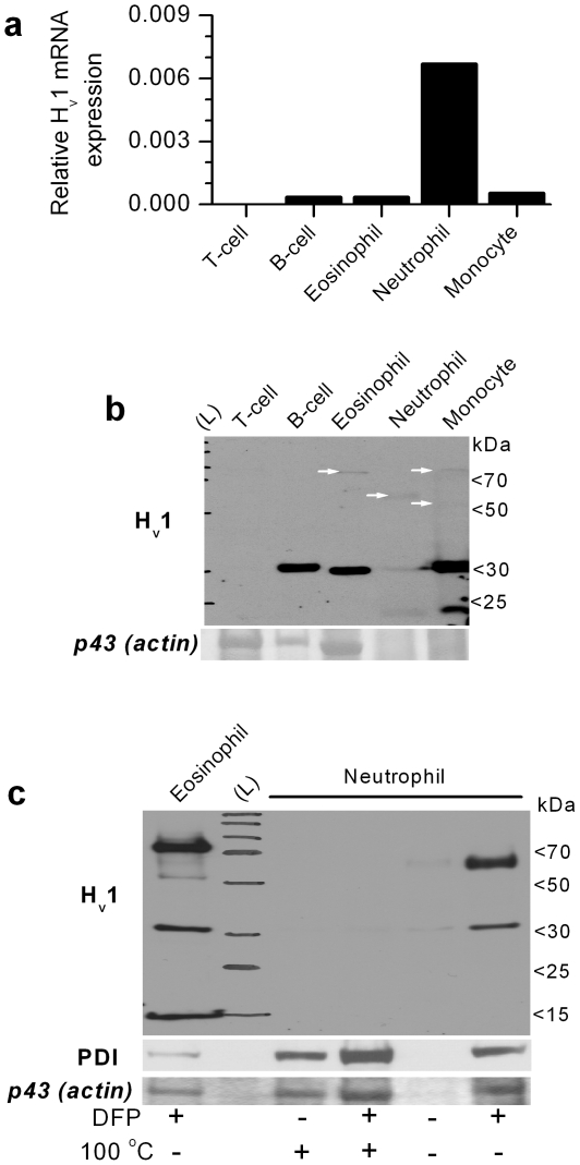
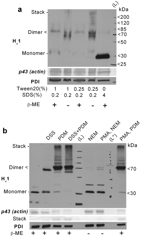
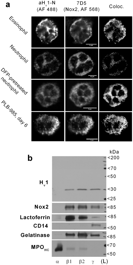
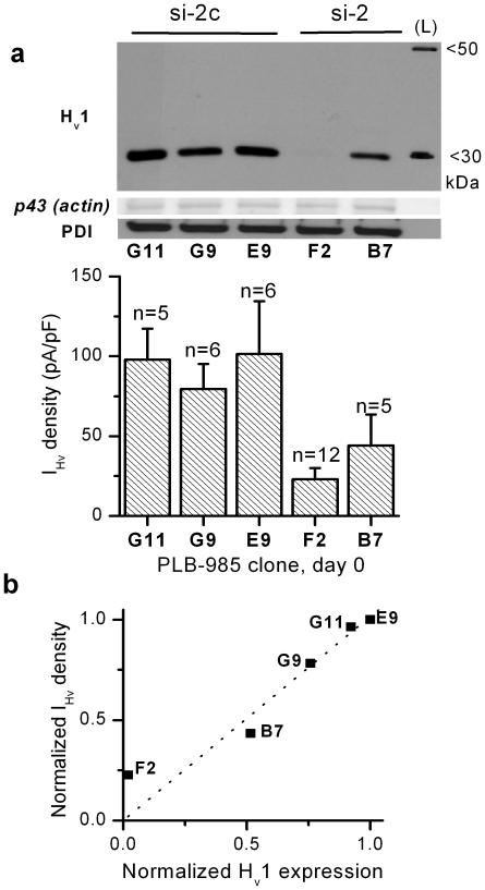
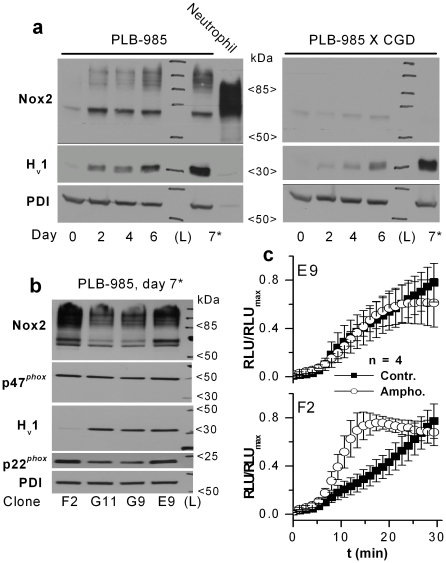
Voltage-gated proton current (I(Hv)) has been characterized in several cell types, but the majority of the data was collected in phagocytes, especially in human granulocytes. The prevailing view about the role of I(Hv) in phagocytes is that it is an essential supporter of the intense and sustained activity of Nox2 (the core enzyme of the phagocyte NADPH oxidase complex) during respiratory burst. Recently H(v)1, a voltage-gated proton channel, was cloned, and leukocytes from H(v)1 knockout mice display impaired respiratory burst. On the other hand, hardly anything is known about H(v)1 in human granulocytes. Using qPCR and a self made antibody, we detected a significant amount of H(v)1 in human eosinophil and neutrophil granulocytes and in PLB-985 leukemia cells. Using different crosslinking agents and detergents in reducing and non-reducing PAGE, significant expression of H(v)1 homodimers, but not that of higher-order multimers, could be detected in granulocytes. Results of subcellular fractionation and confocal imaging indicate that H(v)1 is resident in both plasmalemmal and granular membrane compartments of resting neutrophils. Furthermore, it is also demonstrated that H(v)1 accumulates in phagosome wall during zymosan engulfment together with, but independently of Nox2. During granulocytic differentiation early and parallel upregulation of H(v)1 and Nox2 expression was observed in PLB-985 cells. The upregulation of H(v)1 or Nox2 expression did not require the normal expression of the other molecule. Using RNA interference, we obtained strong correlation between H(v)1 expression and I(Hv) density in PLB-985 cells. It is also demonstrated that a massive reduction in H(v)1 expression can limit the Nox2 mediated superoxide production of PLB-985 granulocytes. In summary, beside monomers native H(v)1 forms stable proton channel dimer in resting and activated human granulocytes. The expression pattern of H(v)1 in granulocytes is optimized to support intense NADPH oxidase activity.


Conflict of interest statement

Competing Interests: The authors have declared that no competing interests exist.

Figures

Figure 1
Figure 1. Expression pattern of Hv1 mRNA and protein in major classes of human peripheral blood leukocytes.
(a) Real-time qPCR analysis of Hv1 mRNA expression relative to cyclophilin mRNA level (similar relative expressions were observed if genes other than cyclophilin were used for normalization). Data represent the mean result of a duplicate experiment. The experiment was repeated in an independent set of leukocytes with similar results. (b) Western blot analysis of Hv1 expression in the same cell types as detected with aHv1-N. Total cell lysates of 106 cells were loaded each lane. The lane with stained molecular weight marker is labeled with (L). Ponceau-stained ∼43 kDa apparent Mw protein band (putative actin) demonstrates the protein load and the quality of the samples. The absence of clear actin band in the neutrophil and monocyte lanes indicates massive protein degradation. Note that immunolabeling of proteins which are considered “house keeping” (e.g. actin or PDI) is of very limited use in case of cell types with very different proteolytic activity, motility and metabolism. Importantly, aHv1-N also detects one or more faint band between ∼60 and ∼80 kDa in myeloid cell types (white arrows). (c) Detection of Hv1 and higher Mw bands is hampered in granulocytes by serine proteases and sample heat treatment. Total cell lysates of 5×10⁵ cells were loaded each lane. Ponceau-stained ∼43 kDa apparent Mw protein band (putative actin) and aPDI labeling demonstrate the degree of protein degradation. Note that anti-PDI signal and the intensity of the actin band is well correlated, which justifies the use of the latter signal to demonstrate protein load and sample quality. DFP incubation for 30 min on ice before cell lysis and heating of the sample (at 100°C for 10 min) were applied as indicated. This experiment was designed based on results from pilot studies, and was performed for demonstration purposes only, thus it was not repeated in this form. In diverse experiments low Mw Hv1 labeling was occasionally observed after DFP treatment, which was independent of the granulocyte type, and likely reflects some remaining protease activity in the given sample.
Figure 2
Figure 2. Hv1 dimers are natively present in human granulocytes.
(a) Total cell lysates of 106 PMNs were loaded each lane. DFP-treated cells were lysed in modified 2x Laemmli sample buffer supplemented with 2 mM PMSF. Detergent composition and the presence of 5% v/v β-mercaptoethanol in the sample buffer are indicated below. (b) WB detection of Hv1 dimers in granulocyte samples treated with amino- (DSS) or thiol-reactive (PDM) crosslinkers or with thiol-reactive alkylating agent (NEM). The increase in labeling with aHv1-N at ∼70 kDa is at the expense of that at ∼30 kDa in cross-linked samples. The appearance of higher (above 75 kDa) Mw aHv1-N-labeled bands is likely, at least in part, due to crosslinking of Hv1 with other proteins. PMA pretreatement (200 nM for 15 min) exerted only minor effect on the vicinal cysteines in the Hv1 dimer. The identity of the faint, ∼40 kDa band is not clear, and it was not consistently detected. Ponceau-stained ∼43 kDa band (putative actin) and near stack region demonstrates the protein load and sample crosslinking. The presence of 5% v/v β-mercaptoethanol in the sample buffer is indicated below. L* denotes unstained Mw marker used for calibrating the routinely used, stained Mw marker.
Figure 3
Figure 3. Partial colocalization of Hv1 and Nox2 in different types of human granulocytes.
(a) Detection of Hv1 (left-most column) and Nox2 (middle) in granulocytes. (Scale bars represent 5 µm). Pretreating neutrophils with DFP improved the detection of Hv1. Colocalization analysis was performed only in cells, in which above-threshold labeling (two times above background, see below) for both proteins could be detected. Colocalizing pixels are displayed as white dots in the most right column. Negligible Alexa Fluor® signals (considered as background) were detected with control primary antibodies (not shown). (b) Western blot analysis of the distribution of Hv1 and Nox2 between granule fractions of resting neutrophils after standard reducing PAGE. Ponceau stain confirmed that similar protein amount was loaded each lane (not shown). The different fractions are: azurophil (α), specific (β1), gelatinase (β2) granules and secretory vesicle together with plasma membrane (γ). Immunodetection of lactoferrin, gelatinase, CD14 and the heavy chain of myeloperoxidase (MPOHC) demonstrates the purity of the membrane preparates .
Figure 4
Figure 4. Partial intracellular colocalization of Hv1 and Nox2 in phagocytosing granulocytes.
Hv1 (first raw) and Nox2 (second raw) tend to cluster during Zymosan phagocytosis, as detected in immunofluorescence experiments using confocal laser microscopy. The locations of zymosans are indicated by white asterisks. Forming (phagocytic cup, first column) and closed phagosomes (second and third column) are visible as round, hollow structures. In further analyses only pixels with intensity at least two times the average intensity (threshold) observed in experiments with control antibodies were included (activated phagocytes displayed significant, diffuse labeling in control experiments, not shown). Clusters of Hv1 and Nox2 are often colocalized (third row). Colocalizing pixels are superimposed as white dots on the dimmed picture of 7D5 labeling (scale bars represent 5 µm). Hv1 clustering is independent of Nox2 in PLB-985 cells (fourth raw). Cell perimeter (as derived form background labeling with Hv1) is outlined in gray, and above-threshold Hv1 labeling is presented as white dots. In the fifth raw above-threshold Hv1 signals are superimposed as white dots over the (dimmed) visible light transmission image of phagocytosing cells. Pseudo color, 3D reconstructions of the cells in the 3rd and 4th columns are provided as supplemental Videos S1 and S2, respectively.
Figure 5
Figure 5. Correlation between Hv1 and voltage-gated proton current.
(a) The expression level of Hv1 (top) and the corresponding IHv density (below) is presented in selected PLB-985 clones. The clones were transfected with a plasmid producing siRNA capable of knocking down Hv1 expression (si-2) or its control siRNA (si-2c). For WB total cell lysates of 106 cells were loaded each lane. The IHv density in clone F2 is significantly smaller than in E9 and G11 (p<0.05, Kruskal-Wallis test) (b) Mean IHv density value is plotted against the corresponding Hv1 signal normalized to p43 signal, as measured with densitometry. The values of each clone are divided by the values of clone E9. Dotted line is the result of linear fit constrained to path through the origin (R>0.97, p<0.005).
Figure 6
Figure 6. The functionally coupled Hv1 and Nox2 are induced in parallel, but largely independently during granulocytic differentiation in PLB-985 cells.
(a) Normal Hv1 expression can be induced in the absence of normal Nox2 level. For WB total cell lysates of 106 cells were loaded each lane. Samples of PLB-985 cells were prepared before (0) and 2, 4, 6 days after inducing differentiation with 0.5% DMFA. In a separate sample (7*) cells were differentiated for 7 days, and DMFA treatment was applied in low-serum culture medium (0.5% v/v) to increase the differentiation pressure. Different anti-Nox2 labeled bands correspond to different glycozilation states of the 65 kDa Nox2 protein. (b) The normal expression of different phox subunits (Nox2, p22phox and p47phox) is not disturbed by strongly reduced Hv1 expression in differentiated PLB-985 cells. (c) Amongst differentiated PLB-985 clones amphotericin B amplifies superoxide production (2.92±0.48 times at 15 min, p<0.05, Mann-Whitney U test) only in clone F2, in which Hv1 expression is strongly diminished. No significant change in superoxide production was detected in E9, G9 and B7 clones in the presence of amphotericin B. Diogenes reagent was used to detect the extracellular release of superoxide. Cells were preincubated for 15 min in a 1∶1 mixture of Diogenes and H-medium with or without 10 µg/ml amphotericin B. At time point 0 cells were activated by 200 nM PMA. Negligible Diogenes luminescence could be detected in PMA-treated, non-differentiated PLB-985 clones and during the preincubation period (not shown).

References

    1. DeCoursey TE. Voltage-gated proton channels and other proton transfer pathways. Physiol Rev. 2003;83:475–579. - PubMed
    1. Sasaki M, Takagi M, Okamura Y. A Voltage Sensor-Domain Protein is a Voltage-Gated Proton Channel. Science. 2006;312:589–592. - PubMed
    1. Ramsey IS, Moran MM, Chong JA, Clapham DE. A voltage-gated proton-selective channel lacking the pore domain. Nature. 2006;440:1213–1216. - PMC - PubMed
    1. Musset B, Cherny VV, Morgan D, Okamura Y, Ramsey IS, et al. Detailed comparison of expressed and native voltage-gated proton channel currents. J Physiol. 2008;586:2477–2486. - PMC - PubMed
    1. Lee SY, Letts JA, Mackinnon R. Functional reconstitution of purified human Hv1 H+ channels. J Mol Biol. 2009;387:1055–1060. - PMC - PubMed

Publication types

MeSH terms