. 2010 Nov 23;5(11):e14102.
doi: 10.1371/journal.pone.0014102.

An Ash2L/RbBP5 heterodimer stimulates the MLL1 methyltransferase activity through coordinated substrate interactions with the MLL1 SET domain

Affiliations

An Ash2L/RbBP5 heterodimer stimulates the MLL1 methyltransferase activity through coordinated substrate interactions with the MLL1 SET domain

Fang Cao et al. PLoS One. .

Abstract

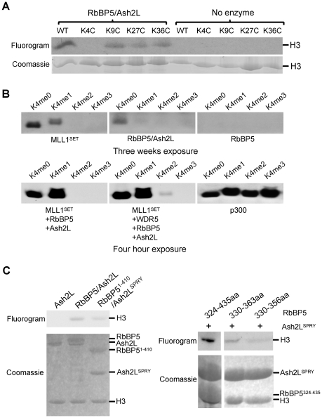
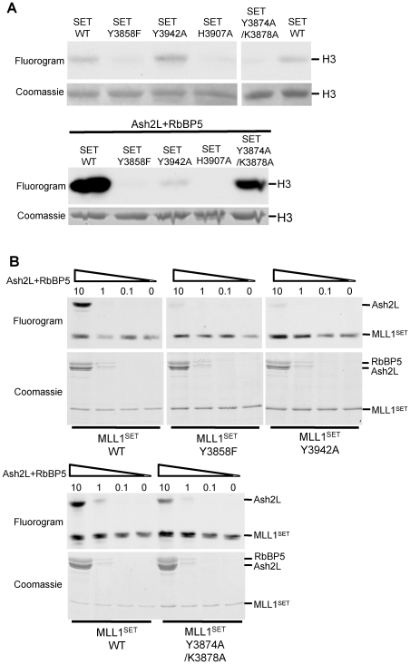
Histone H3 lysine 4 (K4) methylation is a prevalent mark associated with transcription activation and is mainly catalyzed by the MLL/SET1 family histone methyltransferases. A common feature of the mammalian MLL/SET1 complexes is the presence of three core components (RbBP5, Ash2L and WDR5) and a catalytic subunit containing a SET domain. Unlike most other histone lysine methyltransferases, all four proteins are required for efficient H3 K4 methylation. Despite extensive efforts, mechanisms for how three core components regulate MLL/SET1 methyltransferase activity remain elusive. Here we show that a heterodimer of Ash2L and RbBP5 has intrinsic histone methyltransferase activity. This activity requires the highly conserved SPRY domain of Ash2L and a short peptide of RbBP5. We demonstrate that both Ash2L and the MLL1 SET domain are capable of binding to S-adenosyl-L- [methyl-(3)H] methionine in the MLL1 core complex. Mutations in the MLL1 SET domain that fail to support overall H3 K4 methylation also compromise SAM binding by Ash2L. Taken together, our results show that the Ash2L/RbBP5 heterodimer plays a critical role in the overall catalysis of MLL1 mediated H3 K4 methylation. The results we describe here provide mechanistic insights for unique regulation of the MLL1 methyltransferase activity. It suggests that both Ash2L/RbBP5 and the MLL1 SET domain make direct contacts with the substrates and contribute to the formation of a joint catalytic center. Given the shared core configuration among all MLL/SET1 family HMTs, it will be interesting to test whether the mechanism we describe here can be generalized to other MLL/SET1 family members in the future.


Conflict of interest statement

Competing Interests: The authors have declared that no competing interests exist.

Figures

Figure 1
Figure 1. Ash2L/RbBP5 stimulates MLL1 activity in the absence of WDR5.
(A) In vitro HMT assay for MLL1SET and MLL1SET with one, two or three components of the MLL1 complex as indicated on top. For all reactions, 5 µM of MLL1SET was used and all other MLL1 core components were added at equal molar concentration. (B) Scintillation count for in vitro HMT assays using either four-component complex MWAR or three-component mixture MAR as enzymes. Y-axis, scintillation counts (cpm) for the methylation reaction. X-axis, molar concentration of MLL1SET used in different reactions. Other proteins were added at equal molar concentration to MLL1SET in each reaction. All experiments were repeated for three times. The error bar represented standard deviation. (C) In vitro HMT assay using various Ash2L fragments as indicated on top. Top panel, fluorogram of methylated H3. Bottom two panels, Coomassie stained gels for H3 substrate and the MLL1 core components used in the same reaction. (D) In vitro HMT assay using various RbBP5 peptides at equal molar concentration (5 µM) as indicated on top. Top panel, fluorogram of methylated H3. Bottom two panels, Coomassie stained gels for H3 substrate and the MLL1 core components used in the same reaction. Ash2LSPRY and MLL1SET were run at about the same position. RbBP5 peptides were run out of the gel due to their small sizes.
Figure 2
Figure 2. Ash2L/RbBP5 has intrinsic methyltransferase activity.
(A) In vitro HMT assay using ∼5 µM Ash2L/RbBP5 heterodimer as enzymes. Top, fluorogram of methylated H3 after three weeks of exposure. Bottom, Coomassie stained gels for H3 substrate. (B) In vitro HMT assay using 60 µM unmodified, mono-, di- and tri-methylated H3 K4 peptides as substrates. The proteins (∼5 µM final concentration) in each reaction were indicated on bottom. In vitro HAT assay using p300 was included as a quality and loading control for H3 peptides. The exposure times for the top and bottom panels were different and were indicated on bottom. (C) Left, in vitro HMT assay for Ash2L, Ash2L/RbBP5 as well as Ash2LSPRY/RbBP51-410. Right, in vitro HMT assay for Ash2LSPRY and various RbBP5 peptides as indicated on top. Coomassie stained gels for H3 and proteins used in the assays were included on bottom.
Figure 3
Figure 3. Ash2L/RbBP5 binds to substrate SAM.
(A) SAM binding assays for Ash2L/RbBP5 and MLL1SET by UV cross-linking. Left, SAM binding assays for the MLL1 core complex with or without UV cross-linking. Equal molar (∼3 µM) of each MLL1 core component was used. Right, SAM binding by Ash2L and MLL1SET could be chased off by 1000x excess cold SAM (∼0.35 mM) but not by ATP (∼0.35 mM). Black lines indicated expected positions for each protein. 10-fold molar excess of Ash2L/RbBP5 (relative to MLL1SET) was used in this experiment to reduce the exposure time for detecting Ash2L SAM binding. Black lines indicated expected positions for each protein. (B) SAM binding assay for 3 µM equivalent of Ash2L alone or Ash2L/RbBP5 in the presence of increasing amount of MLL1SET. Molar ratios of MLL1SET versus Ash2L/RbBP5 or Ash2L were indicated on top. (C) Image-J quantitation of the results in (B). Y-axis, arbitrary unit for pixel count. (D) Reciprocal SAM binding assays as performed in (B) except ∼3 µM wild type or H3907A MLL1SET was mixed with increasing amount of Ash2L/RbBP5. The molar ratios of Ash2L/RbBP5 versus MLL1SET were indicated on top. (E) Image-J quantitation of the results in (D). Y-axis, arbitrary unit for pixel count. In (B) and (D), Coomassie staining for the same gels was included on bottom. The positions for Ash2L and RbBP5 were indicated on left. *, non-specific protein.
Figure 4
Figure 4. Ash2L/RbBP5 is important for the overall activity of the MLL1 complex.
(A) Co-immunoprecipitation of RbBP5 and Ash2L. Proteins purified through either Flag tag on RbBP5 or RbBP5 mutant (left) or His tag on Ash2L or Ash2L mutant (right) were run on SDS-PAGE for Coomassie staining. (B) In vitro HMT assay for MLL1SET, Ash2L or Ash2L R343A mutant and RbBP5 or RbBP5 mutant as indicated on top. Equal amount of proteins and H3 substrate were used in each reaction. (C) SAM binding assays using proteins indicated on top. Positions of Ash2L and MLL1SET were indicated on right. In (B)-(C), SG refers to RbBP5 F352S/D353G double mutations. Coomassie stained gels were included on bottom as controls. (D) HeLa cells were treated with siRNAs targeting endogenous Ash2L transcripts for 48hrs and then transfected with plasmids expressing Myc-tagged Ash2L or Ash2L mutant R343A. Total proteins were separated on SDS-PAGE and blotted for different antibodies indicated on right. Anti-H3 antibody was used as the loading control.
Figure 5
Figure 5. Ash2L/RbBP5 and MLL1SET coordinate in H3 K4 methylation.
(A) In vitro HMT assays using ∼5 µM of wild type and mutants MLL1SET either alone (top) or with Ash2L/RbBP5 (bottom) as enzymes (B) SAM binding assays for Ash2L and either wild type MLL1SET or SET domain mutants. 3 µM of wild type MLL1SET or SET domain mutants was used in each reaction. Molar ratios of Ash2L/RbBP5 versus MLL1SET were indicated on top. The positions for Ash2L and RbBP5 were indicated on left. Duplicate samples were run for Coomassie staining as the loading controls.
Figure 6
Figure 6. H3 and SAM mediate Ash2L/RbBP5 and MLL1SET interaction.
(A) 150 nM recombinant proteins Flag-Ash2L, His-RbBP5 and His-MLL1SET were mixed in the presence of ∼7.5 µM histone H3, ∼10 µM methylation product AdoHcy or both as indicated on top. After Flag-IP, eluates from M2 agarose beads were separated on SDS-PAGE and immunoblotted for MLL1SET, Ash2L and RbBP5 (indicated on right). 25% input was loaded as the control. (B) “Single catalytic center” model for the regulation of the MLL1 core complex. In this model, Ash2L/RbBP5 and MLL1SET interact with the same H3 substrate and substrate SAM and form a shared active center for catalysis. This structure is further stabilized by WDR5, which simultaneously interacts with RbBP5 and MLL1SET.

References

    1. Dou Y, Milne TA, Tackett AJ, Smith ER, Fukuda A, et al. Physical association and coordinate function of the H3 K4 methyltransferase MLL1 and the H4 K16 acetyltransferase MOF. Cell. 2005;121:873–885. - PubMed
    1. Milne TA, Briggs SD, Brock HW, Martin ME, Gibbs D, et al. MLL targets SET domain methyltransferase activity to Hox gene promoters. Mol Cell. 2002;10:1107–1117. - PubMed
    1. Nakamura T, Mori T, Tada S, Krajewski W, Rozovskaia T, et al. ALL-1 is a histone methyltransferase that assembles a supercomplex of proteins involved in transcriptional regulation. Mol Cell. 2002;10:1119–1128. - PubMed
    1. Guenther MG, Jenner RG, Chevalier B, Nakamura T, Croce CM, et al. Global and Hox-specific roles for the MLL1 methyltransferase. Proc Natl Acad Sci U S A. 2005;102:8603–8608. - PMC - PubMed
    1. Ruthenburg AJ, Allis CD, Wysocka J. Methylation of lysine 4 on histone H3: intricacy of writing and reading a single epigenetic mark. Mol Cell. 2007;25:15–30. - PubMed

Publication types

MeSH terms