. 2010 Nov 19;5(11):e14056.
doi: 10.1371/journal.pone.0014056.

The preferred substrates for transglutaminase 2 in a complex wheat gluten digest are Peptide fragments harboring celiac disease T-cell epitopes

Affiliations

The preferred substrates for transglutaminase 2 in a complex wheat gluten digest are Peptide fragments harboring celiac disease T-cell epitopes

Siri Dørum et al. PLoS One. .

Abstract

Background: Celiac disease is a T-cell mediated chronic inflammatory disorder of the gut that is induced by dietary exposure to gluten proteins. CD4+ T cells of the intestinal lesion recognize gluten peptides in the context of HLA-DQ2.5 or HLA-DQ8 and the gluten derived peptides become better T-cell antigens after deamidation catalyzed by the enzyme transglutaminase 2 (TG2). In this study we aimed to identify the preferred peptide substrates of TG2 in a heterogeneous proteolytic digest of whole wheat gluten.

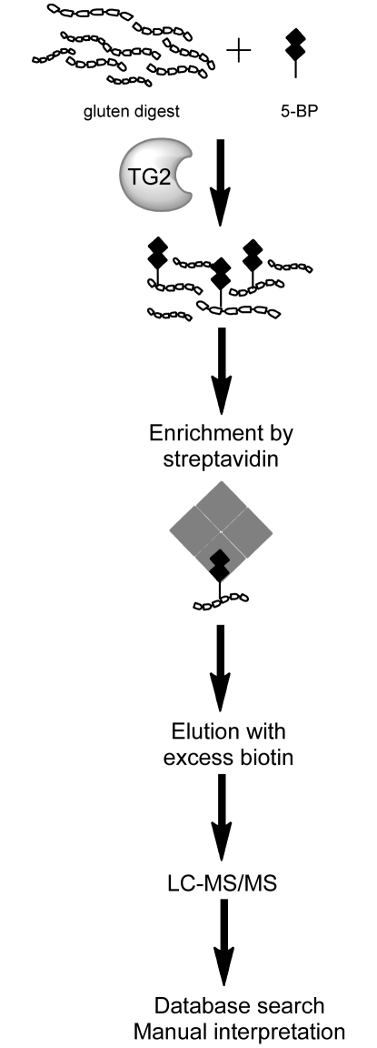
Methods: A method was established to enrich for preferred TG2 substrates in a complex gluten peptide mixture by tagging with 5-biotinamido-pentylamine. Tagged peptides were isolated and then identified by nano-liquid chromatography online-coupled to tandem mass spectrometry, database searching and final manual data validation.

Results: We identified 31 different peptides as preferred substrates of TG2. Strikingly, the majority of these peptides were harboring known gluten T-cell epitopes. Five TG2 peptide substrates that were predicted to bind to HLA-DQ2.5 did not contain previously characterized sequences of T-cell epitopes. Two of these peptides elicited T-cell responses when tested for recognition by intestinal T-cell lines of celiac disease patients, and thus they contain novel candidate T-cell epitopes. We also found that the intact 9mer core sequences of the respective epitopes were not present in all peptide substrates. Interestingly, those epitopes that were represented by intact forms were frequently recognized by T cells in celiac disease patients, whereas those that were present in truncated versions were infrequently recognized.

Conclusion: TG2 as well as gastrointestinal proteolysis play important roles in the selection of gluten T-cell epitopes in celiac disease.


Conflict of interest statement

Competing Interests: The authors have declared that no competing interests exist.

Figures

Figure 1
Figure 1. Schematic view of the established method to enrich and analyze TG2 peptide substrates.
A PTCEC digest of wheat gluten was mixed with a small amount of 5-BP which served as a substrate for TG2 in a transamidation reaction. The transamidated, biotinylated peptides were enriched from the digest using magnetic streptavidin beads, eluted with an excess of biotin and analyzed by LC-MS/MS. Database searching was performed using a database made up of all entries of Triticum aestivum present in the Uniprot database. In addition, MS/MS spectra were manually inspected.
Figure 2
Figure 2. Specific enrichment of transamidated peptides.
A PTCEC digest of whole gluten was spiked with the transamidated DQ2-α-I and DQ2-γ-II peptides. MALDI-TOF spectra before (upper panel) and after enrichment (lower panel) are shown. The inset shows the four signals obtained for each of the two enriched peptides that correspond to the transamidated peptide, its sodium adduct, its potassium adduct and the adduct with two sodium ions, respectively. All signals observed in the lower mass range (up to m/z 900) were derived from matrix clusters.
Figure 3
Figure 3. T-cell recognition of gluten peptides #19 and #25.
The identified TG2 peptide substrates VPVPQLQPQNPSQQQPQEQVPL (peptide #9), PHQPQQQVPQPQQPQQPF (peptide #19), SHQQQPFPQQPYPQQPYPS (peptide #25) and SFPQPQPQQPQQPS (peptide #30) were tested together with a panel of synthetic epitope peptides for recognition by three T-cell lines, TCL.BW.CD-E (A), TCL.497.C.1.3 (B), and TCL.422.02.2.4 (C), generated from biopsies of HLA-DQ2.5-positive celiac disease patients. The sequences of the other synthetic peptides tested were|: DQ2-α-I, QLQPFPQPELPY; DQ2-α-II, PQPELPYPQPQLPY; α33mer, LQLQPFPQPELPYPQPELPYPQPELPYPQPQPF; DQ2-γ-I, pyroglutamic acid-PQQPQQSFPEQQRP (in A), pyroglutamic acid-PEQPQQSFPEQERP (in B and C); DQ2-γ-II, GIIQPEQPAQL; DQ2-γ-III, FPEQPEQPYPEQ (in A), FPQQPEQPYPQQ (in B and C); DQ2-γ-IV, FSQPEQEFPQPQ; DQ2-γ-VI, PEQPFPEQPEQ; DQ2-γ-VIIa, TEQPEQPFPQP; DQ2-γ-VIIb, FPQPEQEFPQPQ; DQ2-γ-III/DQ2-γ-VI, LQPEQPFPEQPEQPYPEQPQ; γ23mer, EQPFPEQPEQPYPEQPEQPFPQP; ω17mer, QPQQPFPQPEQPFPWQP. Note that peptides #25 and #19 were tested at different concentrations. The α33mer peptide was tested at 2 µM while the other peptides were tested at 10 µM. Responses were measured in a proliferation assay by the incorporation of 3H-thymidine (counts per minute (CPM)×103). The dashed line indicates two-fold background proliferation observed with medium only. Peptides #25 and #19 were tested in triplicates; other antigens were tested in duplicates. Error bars indicate the standard error of mean. The experiment was repeated twice. The HLA-restriction of the T-cell line TCL.422.02.2.4 was assessed by blocking recognition of peptide #25 using anti-DR, anti-DQ and anti-DP specific monoclonal antibodies (D). Similar results were obtained for T-cell line TCL.497.C.1.3.

Similar articles

Cited by

References

    1. Sollid LM. Molecular basis of celiac disease. Annu Rev Immunol. 2000;18:53–81. - PubMed
    1. Lundin KE, Scott H, Hansen T, Paulsen G, Halstensen TS, et al. Gliadin-specific, HLA-DQ(alpha 1*0501,beta 1*0201) restricted T cells isolated from the small intestinal mucosa of celiac disease patients. J Exp Med. 1993;178:187–196. - PMC - PubMed
    1. Molberg O, Kett K, Scott H, Thorsby E, Sollid LM, et al. Gliadin specific, HLA DQ2-restricted T cells are commonly found in small intestinal biopsies from coeliac disease patients, but not from controls. Scand J Immunol. 1997;46:103–109. - PubMed
    1. Johansen BH, Vartdal F, Eriksen JA, Thorsby E, Sollid LM. Identification of a putative motif for binding of peptides to HLA-DQ2. Int Immunol. 1996;8:177–182. - PubMed
    1. van der Wal Y, Kooy YM, Drijfhout JW, Amons R, Koning F. Peptide binding characteristics of the coeliac disease-associated DQ(alpha1*0501, beta1*0201) molecule. Immunogenetics. 1996;44:246–253. - PubMed

Publication types

MeSH terms