. 2010 Nov 18;6(11):e1001214.
doi: 10.1371/journal.pgen.1001214.

The parental non-equivalence of imprinting control regions during mammalian development and evolution

Affiliations

The parental non-equivalence of imprinting control regions during mammalian development and evolution

Reiner Schulz et al. PLoS Genet. .

Abstract

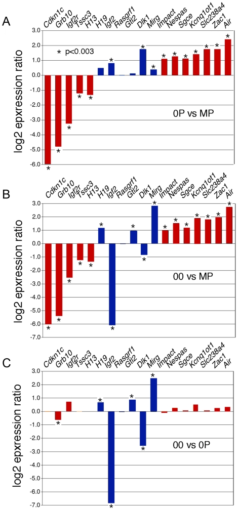
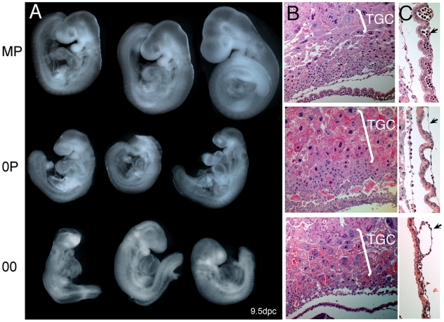
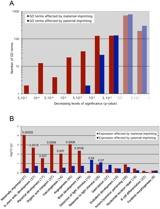
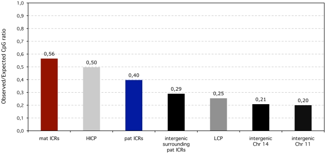
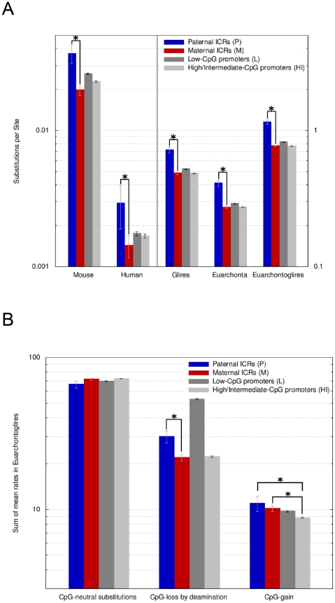
In mammals, imprinted gene expression results from the sex-specific methylation of imprinted control regions (ICRs) in the parental germlines. Imprinting is linked to therian reproduction, that is, the placenta and imprinting emerged at roughly the same time and potentially co-evolved. We assessed the transcriptome-wide and ontology effect of maternally versus paternally methylated ICRs at the developmental stage of setting of the chorioallantoic placenta in the mouse (8.5dpc), using two models of imprinting deficiency including completely imprint-free embryos. Paternal and maternal imprints have a similar quantitative impact on the embryonic transcriptome. However, transcriptional effects of maternal ICRs are qualitatively focused on the fetal-maternal interface, while paternal ICRs weakly affect non-convergent biological processes, with little consequence for viability at 8.5dpc. Moreover, genes regulated by maternal ICRs indirectly influence genes regulated by paternal ICRs, while the reverse is not observed. The functional dominance of maternal imprints over early embryonic development is potentially linked to selection pressures favoring methylation-dependent control of maternal over paternal ICRs. We previously hypothesized that the different methylation histories of ICRs in the maternal versus the paternal germlines may have put paternal ICRs under higher mutational pressure to lose CpGs by deamination. Using comparative genomics of 17 extant mammalian species, we show here that, while ICRs in general have been constrained to maintain more CpGs than non-imprinted sequences, the rate of CpG loss at paternal ICRs has indeed been higher than at maternal ICRs during evolution. In fact, maternal ICRs, which have the characteristics of CpG-rich promoters, have gained CpGs compared to non-imprinted CpG-rich promoters. Thus, the numerical and, during early embryonic development, functional dominance of maternal ICRs can be explained as the consequence of two orthogonal evolutionary forces: pressure to tightly regulate genes affecting the fetal-maternal interface and pressure to avoid the mutagenic environment of the paternal germline.


Conflict of interest statement

The authors have declared that no competing interests exist.

Figures

Figure 1
Figure 1. Methylation patterns in maternal-imprint free 0P and complete-imprint free 00 embryos at 8.5dpc.
A, Bisulfite genomic sequencing of ICRs associated with the H19 and Kcnq1ot1 loci. The paternally methylated H19 ICR was methylated in wildtype (MP) and in maternal imprint-free (0P) but unmethylated in imprint-free (00) visceral yolk sacs. The maternal Kcnq1ot1 ICR was hypomethylated in both 0P and 00 material, in agreement with the lack of maternal imprints in these embryos. Nucleotide positions are reported in reference to the gene transcription start (+1). B, Normal methylation of LINE1 and IAP retrotransposons in MP, 0P, and 00 8.5dpc embryos as established by DNA blot hybridization after cleavage with the methylation-insensitive restriction endonuclease MspI or the methylation sensitive isoschizomer HpaII.
Figure 2
Figure 2. Expression abnormalities at imprinted loci in 8.5dpc 0P and 00 embryos.
Microarray-measured gene expression ratios for known imprinted genes under the control of either maternally (red) or paternally (blue) methylated ICRs. The absolute expression levels were calculated using GC-RMA. Asterisks indicate a significant difference in expression (GCOS p-value <0.003). A, Imprinted gene expression in maternal imprint-free 0P embryos relative to wildtype biparental MP embryos. Maternally repressed imprinted genes (Airn, Zac1…) were upregulated, while maternally expressed imprinted genes (Cdkn1c, Grb10…) were downregulated. Changes observed at the paternally imprinted H19, Igf2 and Dlk1 genes are potentially secondary to the upregulation of Zac1, which is known to regulate the expression of multiple genes acting within the same functional network . B, Imprinted gene expression in 00 imprint-free relative to MP embryos. As expected, maternally imprinted genes are as affected in 00 and 0P embryos compared to MP embryos. Genes controlled by paternal ICRs show changes expected to occur in the absence of paternal imprints, i.e. an upregulation of H19 and Gtl2 and subsequent downregulation of Igf2 and Dlk1. C, In this comparison of 00 and 0P embryos, the significant changes in the expression of paternally imprinted genes persist, while for most maternally imprinted genes, no significant differential expression is observed, as is expected since both samples lack maternal germline imprints.
Figure 3
Figure 3. Net effects of loss of paternal and maternal germline imprints on early mouse development.
A, Gross morphology of 9dpc MP, 0P, and 00 embryos. Note that 0P and 00 embryos are very similar in size and phenotype, which implies a minor contribution of paternal imprints to early development. Especially notable signs are the intrauterine growth retardation, open anterior neural tube, enlarged pericardium, reduced head size and abnormal craniofacial features. B., Hyperplasia of the trophoblast giant cell layer (TGC) in 0P and 00 conceptuses. C., Severe deficiency in the vascularization (arrows) of 0P and 00 visceral yolk sacs. Note that erythrocytes are present in MP but are largely absent in 0P and 00 VYS.
Figure 4
Figure 4. Maternal versus paternal imprint influence on specific molecular functions and developmental processes.
Overrepresentation analysis results for Gene Ontology (GO) categories. The analysis was carried out separately and independently for two different scoring schemes. Essentially, the “maternal” scheme assigned a non-zero score if the gene's expression pattern across the MP, 0P and 00 samples was consistent with the gene's expression being affected by maternal but not paternal germline imprints (red bars). Analogously, the “paternal” scoring scheme gave non-zero scores to genes that appeared affected by paternal but not maternal germline imprints (blue bars). A, p-value distribution across all GO categories for genes affected by maternal versus paternal imprints. P-values below 0.1 were considered as significant, greater values do not indicate relevant impact (shaded bars). The y-axis shows the –log10 value of the GO terms numbers. Note the skewed distribution of paternal-dependent GO terms towards the less significant p-values >0.001. On the contrary, maternal-dependent GO terms are highly clustered on the left towards p <0.001, indicating a stronger biological impact of maternal imprints. B, Loss of maternal imprints affects specific early development pathways essential for embryonic viability (in utero development, placentation, vasculogenesis, solute transport), while paternal imprints have small effects. The custom Maternally imprinted category served as a control: it was significantly overrepresented (p = 0.00005) under the maternal but not the paternal scoring scheme. The y-axis shows the –log10 value of the multiple testing-corrected p-value. The absolute p-values with high confidence scores (<0.1) are reported on top of the corresponding bars. The number of genes present in each biological category is shown in brackets.
Figure 5
Figure 5. Observed over expected CpG ratios of ICRS compared to non-imprinted promoters and intergenic regions in the human lineage.
In terms of CpG content, maternal (mat) ICRs are similar to High to Intermediate CpG content promoters (HICP) (0.56 versus 0.5). Despite being in intergenic regions, the CpG content of paternal (pat) ICRs are higher than for intergenic regions in general (measured along Chromosomes 11 and 14) and also than surrounding intergenic regions. Low (LCP) CpG-content promoters are similar to intergenic regions.
Figure 6
Figure 6. Paternal ICRs have evolved faster than maternal ICRs and have endured a higher rate of CpG loss by deamination.
A, Overall amount of nucleotide changes at paternal and maternal ICRs. Amount of change is expressed along the y-axis (log10 scale) as the sum of the Ambiore-estimated branch lengths for singleton branches (human, mouse), for the respective sub-tree (euarchonta, glires) or for the entire tree (euarchontoglires). Sequence categories are: P =  paternal ICRs, M =  maternal ICRs, L =  Low CpG-content promoters, HI  =  High and Intermediate CpG-content promoters. Error bars are 95% confidence intervals. Significant (non-overlapping 95% confidence intervals) changes of interest are labelled with asterisks. Paternal ICRs present with the most evolutionary change, compared to all other sequence types and to maternal ICRs in particular. The values for human and mouse are two orders of magnitude lower than for euarchontoglires, euarchonta and glires. Hence, the use of two y-axis scales (left versus right). B, Rates of substitutions occurring at CpG dinucleotides in euarchontoglires. The estimated substitution rates are relative to each category's overall rate of evolution, e.g., the fact that paternal ICRs are fast evolving intergenic regions, while all other categories are promoter-associated, has been normalized for. Nevertheless, the rate of CpG-loss by deamination at paternal ICRs is higher than for maternal ICRs. Maternal ICRs loose CpGs at the same pace as HI promoters but gain CpGs at a faster rate.

Comment in

  • Methylation from mother.
    Muers M. Muers M. Nat Rev Genet. 2011 Jan;12(1):6. doi: 10.1038/nrg2923. Epub 2010 Dec 7. Nat Rev Genet. 2011. PMID: 21135864 No abstract available.

References

    1. Wood AJ, Oakey RJ. Genomic imprinting in mammals: emerging themes and established theories. PLoS Genet. 2006;2:e147. doi: 10.1371/journal.pgen.0020147. - DOI - PMC - PubMed
    1. Tucker KL, Beard C, Dausmann J, Jackson-Grusby L, Laird PW, et al. Germ-line passage is required for establishment of methylation and expression patterns of imprinted but not of nonimprinted genes. Genes Dev. 1996;10:1008–1020. - PubMed
    1. Bourc'his D, Xu GL, Lin CS, Bollman B, Bestor TH. Dnmt3L and the establishment of maternal genomic imprints. Science. 2001;294:2536–2539. - PubMed
    1. Kaneda M, Okano M, Hata K, Sado T, Tsujimoto N, et al. Essential role for de novo DNA methyltransferase Dnmt3a in paternal and maternal imprinting. Nature. 2004;429:900–903. - PubMed
    1. Bourc'his D, Bestor TH. Origins of extreme sexual dimorphism in genomic imprinting. Cytogenet Genome Res. 2006;113:36–40. - PubMed

Publication types

LinkOut - more resources