. 2010 Nov 18;5(11):e14050.
doi: 10.1371/journal.pone.0014050.

Photounbinding of calmodulin from a family of CaM binding peptides

Affiliations

Photounbinding of calmodulin from a family of CaM binding peptides

Klaus G Neumüller et al. PLoS One. .

Abstract

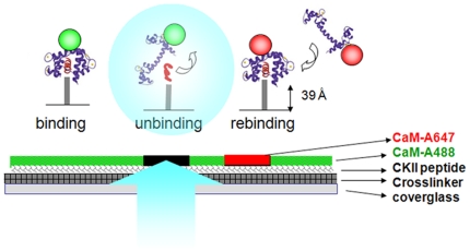
Background: Recent studies have shown that fluorescently labeled antibodies can be dissociated from their antigen by illumination with laser light. The mechanism responsible for the photounbinding effect, however, remains elusive. Here, we give important insights into the mechanism of photounbinding and show that the effect is not restricted to antibody/antigen binding.

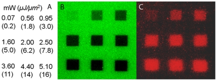
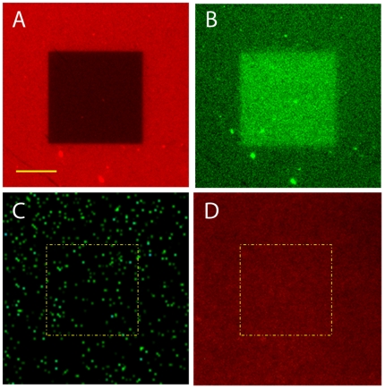
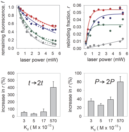
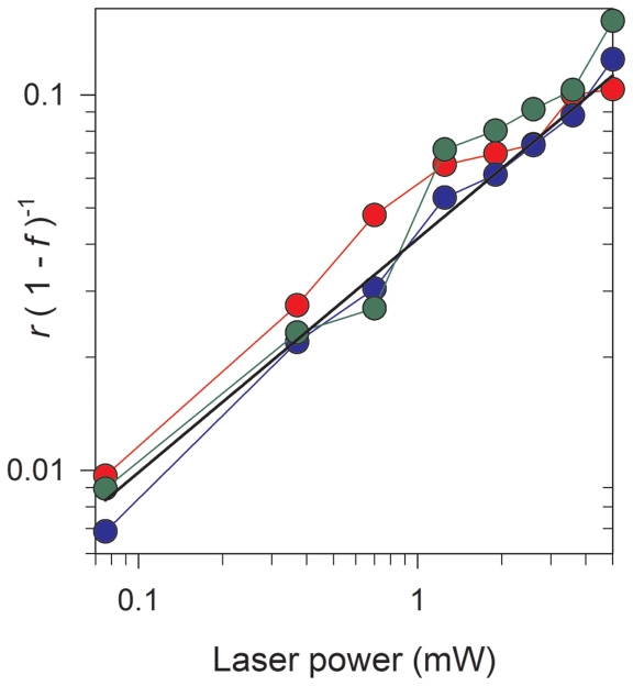
Methodology/principal findings: We present studies of the photounbinding of labeled calmodulin (CaM) from a set of CaM-binding peptides with different affinities to CaM after one- and two-photon excitation. We found that the photounbinding effect becomes stronger with increasing binding affinity. Our observation that photounbinding can be influenced by using free radical scavengers, that it does not occur with either unlabeled protein or non-fluorescent quencher dyes, and that it becomes evident shortly after or with photobleaching suggest that photounbinding and photobleaching are closely linked.

Conclusions/significance: The experimental results exclude surface effects, or heating by laser irradiation as potential causes of photounbinding. Our data suggest that free radicals formed through photobleaching may cause a conformational change of the CaM which lowers their binding affinity with the peptide or its respective binding partner.


Conflict of interest statement

Competing Interests: The authors have declared that no competing interests exist.

Figures

Figure 1
Figure 1. Schematic of the photounbinding assay and sample preparation.
CKII peptides were attached to a glass surface via an SM(PEG)8 crosslinker followed by CaM-A488 incubation. After light illumination to induce photounbinding of the CKII peptide/CaM-488 complexes, the surface was re-incubated with CaM-A647 to visualize free binding sites in the previously illuminated regions.
Figure 2
Figure 2. Unbinding of CaM-A488 and CKII(290-312) peptide by 488 nm laser light.
A: laser power and intensity used to illuminate the corresponding patches in B: ‘bleaching’ pattern (CaM-A488 fluorescence, scale bar: 20 µm), and C: rebinding pattern (CaM-A647 fluorescence).
Figure 3
Figure 3. Photounbinding occurs for labeled, but not for unlabeled CaM.
A: Illuminated patch of CaM-A647 and CKII(290–312) by 633 nm laser light; laser power: 190 µW (flux  = 589 nJ/µm2), scale bar  = 10 µm. B: rebinding pattern (CaM-A488 fluorescence). C: illumination of unlabeled CaM and CKII(290–312) peptide by 488 nm laser light within the indicated patch (yellow dashed line); laser power: 370 µW (flux  = 1.15 µJ/µm2), green dots: fluorescent beads to allow proper focusing. D: no rebinding of A647 was observed after laser illumination within the corresponding patch.
Figure 4
Figure 4. Photounbinding is dependent on the initial dissociation constant of the molecular system.
Remaining Fluorescence (A) and corresponding rebinding (B) at various laser powers for peptides CKII(290–312) (grey symbols), CKII(292–312) (green symbols), CKII(293–312) (blue symbols), and CKII(294–312) (red symbols). A: single exponential (solid line) and double exponential (dotted line) fits to the unbinding data. B: single rising-exponential fits to the rebinding data. C: summary of maximal photounbinding values for all tested peptides after 1PE laser illumination (λexc = 488 nm; P = 3.6 mW) and one scan iteration (solid bars) and two scan iteration (open bars) in comparison. D: photounbinding threshold decreases for the lower affinity peptides (Kd: 3–570×10−13 M) the graph shows the relative increase of rebinding when photounbinding laser power is doubled to 7.2 mW. Uncertainties for the rebinding fraction and remaining fluorescence fraction due to variablilty in CKII-CaM coatings and alignment of the coverglasses are less than 15% for each data point, whereas those associated with the laser power are negligible.
Figure 5
Figure 5. Plot of the rebinding to fluorescence-loss ration [r/(1−f)] as a function of laser power for a single line scan.
Data is shown for peptides CKII(290–312) (grey symbols), CKII(292–312) (green symbols), CKII(293–312) (blue symbols), and CKII(294–312) (red symbols); The plot shows that the rebinding fraction is not directly proportional to the loss of fluorescence, but is suppressed at lower laser powers. The solid black line is a least-square fitted power-law to the CKII(293–312) peptide data (blue symbols) and given by: r/(1–f) = 0.04 P 0.6, where P is the laser power.
Figure 6
Figure 6. Photounbinding of labeled phalloidin from actin filaments.
A: Ph-488 fluorescence after illumination at 488 nm (1PE) and 20 µW (62.0 nJ/µm2) (bleached patch is indicated in yellow); B: rebinding of Ph-647 within the previously illuminated area; C: photounbinding in a human fibrosarcoma cell, three squares were bleached (2PE, 800 nm) with different laser intensities left: 14 mW (flux  = 10.7 mJ/µm2); top: 20 mW (15.4 mJ/µm2); right: 24 mW (18.4 mJ/µm2).
Figure 7
Figure 7. Photounbinding using the chemical fluorescence stabilizer ascorbic acid.
A: Remaining CaM-A488 fluorescence (open symbols) and the corresponding rebinding (solid symbols) after two-photon excitation (Ti:Sa laser λexc: 800 nm) with the addition of 8 mM ascorbic acid (squares) and without (circles). Photobleaching (and photounbinding) is partly prevented by the stabilizer as expected. B: Control study with A488 fluorophores directly covalently bound to the SM-PEG8 crosslinker via a tripeptide (H-Gly-Gly-Cys-OH). As expected the Alexa 488 fluorescence was stabilized to a comparable extent in presence of ascorbic acid (squares), however no photounbinding was detected. The two data sets have been fitted with a (2 parameter) single exponential function. Uncertainties for the rebinding fraction and remaining fluorescence fraction due to variablilty in CKII-CaM coatings and alignment of the coverglasses are less than 15% for each data point, whereas those associated with the laser power are negligible.
Figure 8
Figure 8. Jablonski energy diagram for the formation of radicals through bleaching of a generic flourophore (S).
ka,: Excitation rate; kd: total (radiative & non-radiative) decay rate of the fluorophore from the excited- to the ground- singlet state; kisc: intersystem crossing rate; kbs* & kbt*: Bleaching rates of the excited singlet & triplet states into excited bleached (dark) states via radicalization of a surrounding molecule (X→X* or X'→X'*).

References

    1. Bingaman S, Huxley VH, Rolando ER. Fluorescent Dyes Modify Properties of Proteins Used in Microvascular Research. Microcirculation. 2003;10(2):221–231. - PubMed
    1. Luschtinetz F, Dosche C, Kumke MU. Influence of Streptavidin on the Absorption and Fluorescence Properties of Cyanine Dyes. Bioconjugate Chem. 2009;20(3):576–582. - PubMed
    1. Hoebe RA, Van Der Voort HTM, Stap J, Van Noorden CJF, Manders EMM. Qantitative Determination of the Reduction of Phototoxicity and Photobleaching by Controlled Light Exposure Microscopy. J Microsc. 2008;231(Pt 1):9–20. - PubMed
    1. Dobrucki JW, Feret D, Noatynsky A. Scattering of Exciting Light by Live Cells in Fluorescence Confocal Imaging: Phototoxic Effects and Relevance for FRAP Studies. Biophys J. 2007;93:1778–1786. - PMC - PubMed
    1. Remington SJ. Fluorescent Proteins: Maturation, Photochemistry and Photophysics. Curr. Opinion in Struct Biol. 2006;16:714–721. - PubMed

Publication types

MeSH terms