. 2010 Nov 18;6(11):e1001197.
doi: 10.1371/journal.ppat.1001197.

TGF-b2 induction regulates invasiveness of Theileria-transformed leukocytes and disease susceptibility

Affiliations

TGF-b2 induction regulates invasiveness of Theileria-transformed leukocytes and disease susceptibility

Marie Chaussepied et al. PLoS Pathog. .

Abstract

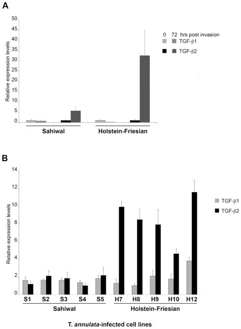
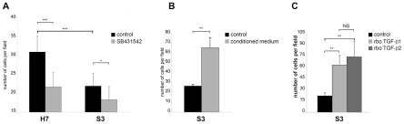
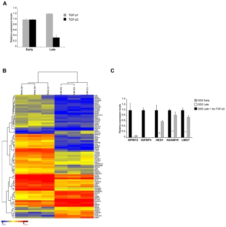
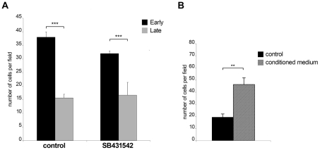
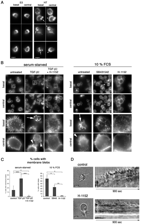
Theileria parasites invade and transform bovine leukocytes causing either East Coast fever (T. parva), or tropical theileriosis (T. annulata). Susceptible animals usually die within weeks of infection, but indigenous infected cattle show markedly reduced pathology, suggesting that host genetic factors may cause disease susceptibility. Attenuated live vaccines are widely used to control tropical theileriosis and attenuation is associated with reduced invasiveness of infected macrophages in vitro. Disease pathogenesis is therefore linked to aggressive invasiveness, rather than uncontrolled proliferation of Theileria-infected leukocytes. We show that the invasive potential of Theileria-transformed leukocytes involves TGF-b signalling. Attenuated live vaccine lines express reduced TGF-b2 and their invasiveness can be rescued with exogenous TGF-b. Importantly, infected macrophages from disease susceptible Holstein-Friesian (HF) cows express more TGF-b2 and traverse Matrigel with great efficiency compared to those from disease-resistant Sahiwal cattle. Thus, TGF-b2 levels correlate with disease susceptibility. Using fluorescence and time-lapse video microscopy we show that Theileria-infected, disease-susceptible HF macrophages exhibit increased actin dynamics in their lamellipodia and podosomal adhesion structures and develop more membrane blebs. TGF-b2-associated invasiveness in HF macrophages has a transcription-independent element that relies on cytoskeleton remodelling via activation of Rho kinase (ROCK). We propose that a TGF-b autocrine loop confers an amoeboid-like motility on Theileria-infected leukocytes, which combines with MMP-dependent motility to drive invasiveness and virulence.


Conflict of interest statement

The authors have declared that no competing interests exist.

Figures

Figure 1
Figure 1. Induction of TGF-b2 by T. annulata is greater in infected macrophages isolated from disease-susceptible compared to disease-resistant bovine hosts.
A, Macrophages isolated from Sahiwal and HF cows were infected in vitro with sporozoites of the Hissar strain of Theileria annulata. RNA was isolated immediately prior to infection (0h) and 72h post-infection and the relative amounts of TGF-b1 and TGF-b2 transcript measured by real-time RT-PCR. B, Total RNA was extracted from T. annulata-infected cell lines obtained from infection of Sahiwals (S1–5) and HF (H7–12) animals. TGF-b1 and TGF-b2 relative mRNA levels were determined by real-time RT-PCR. The relative value obtained for the cell line showing the lowest expression of each cytokine (H8 for TGF-b1 and S4 for TGF-b2) was used for comparison and arbitrary set at 1. Error bars represent standard deviations for triplicate samples. No significant difference between Sahiwal and Holstein-Friesian cells was found for TGF-b1 (p = 0.710), in contrast to TGF-b2 that was 7-fold upregulated (p<0.001) in all 5 H cells lines.
Figure 2
Figure 2. The invasive capacity of Theileria-transformed HF macrophages is greater than Sahiwal-infected macrophages and is TGF-b-dependent.
A, The invasive capacity of H7 and S3 cells was determined by Matrigel chamber assays. Seven independent experiments were performed and each sample counted three times. The y-axis is the number of migrated cells for 10 independent fields. B, Conditioned medium from H7 cultured cells stimulates the invasive capability of S3 cells (*** p<0.0005). Invasion assay was performed on untreated S3 cells and S3 cells that had been incubated for 24h prior to the assay with culture supernatants from H7 cells grown overnight in low-serum (0.5% FCS) containing media. C, Addition of recombinant TGF-b1 and TGF-b2 leads to an increase in the invasive index of Theileria-infected S3 macrophages (** p<0.005).
Figure 3
Figure 3. In vitro attenuation is associated with lower TGF-b2 expression and altered transcripts levels of TGF-b-regulated genes.
A, Total RNA was extracted from early and late passage ODE cells. TGF-b1 and TGF-b2 relative mRNA levels were determined by real-time RT-PCR. B, Total RNA from early and late passage ODE cells (3 replicates each) was hybridised to a microarray representing known bovine genes, including 1,158 targets of TGF-b. 76 of these genes were identified as differentially expressed using the criteria of a false discovery rate <5% (Rank Product analysis) and a fold change >2. Hierarchical clustering was performed on this gene set and the results are depicted as a heat-map. The gene symbol is shown on the right of the heat-map, where colour is used to represent gene expression level: blue (low), yellow (intermediate) and red (high). C, Total RNA was extracted from early and late passage ODE cells, as well as late passage ODE cells treated for 24h with rboTGF-b2 (5ng/ml). Relative mRNA levels for the indicated genes were determined by real-time RT-PCR and normalized to HPRT1 relative levels.
Figure 4
Figure 4. Invasiveness is reduced upon in vitro attenuation of host cell virulence.
A, Early passage Ode cells display higher invasive capability compared to late passage Ode (***p<0.005). The invasive capacity of early passage Ode is reduced (***p<0.005) upon TGF-R blockade by SB431542 B, Culture supernatant from early passage ODE cells stimulates the invasive capability of late passage ODE cells (**p<0.005). Invasion assay was performed on untreated late passage ODE cells and late passage cells that had been incubated for 24h prior to the assay with culture supernatants from early passage ODE cells grown overnight in low-serum (0.5% FCS) containing media.
Figure 5
Figure 5. TGF-b promotes podosomal adhesion structures and ROCK-mediated cortical actin-rich membrane blebs.
A, S3 and H7 cells were seeded onto a gelatin/fibronectin matrix. After 24h in culture, the actin cytoskeleton was visualized by IF microscopy. Basal and central focal planes are shown and actin structures were visualized by phalloidin staining at basal adhesion sites and in the lateral cortex, respectively. B, Left panel: H7 cells were serum starved in 0.5% FCS for 24h and then seeded onto a gelatin/fibronectin matrix in starvation medium and in the absence or presence of TGF-b2 (10ng/ml) and H-1152 (10uM), as indicated. Right panel: H7 cells were grown in complete growth medium and then seeded onto a gelatin/fibronectin matrix in complete growth medium (10% FCS) and in the absence or presence of SB431542 and H-1152, as indicated. Analyses was performed as described in A. Magnifications in lower half of the panels are four-fold of areas highlighted with frames. C, Statistical analysis of experiment shown in panel B. Percentages of cells with membrane blebs within total cell populations was determined. Every cell in at least four independent areas per condition was analysed. 0.5% FCS: control: n = 121 (15.11%−/+9.7), TGF-b: n = 235 (42.00%−/+8.5), TGF-b+H-1152 N = 143 (1%); 10% FCS: control: n = 60 (69.27%−/+12.12), SB431542 = SB43: n = 167 (35.84%−/+6.94), H-1152: n = 81 (1.66%−/+1.47). Arrows indicate podosomal adhesion structures, arrowheads membrane blebs. D, Infected cells were embedded in fibrillar collagen and live-cell video microscopy was performed 24h after embedding. Where indicated, 10uM Rho-kinase inhibitor H-1152 was added 1h before acquisition of movies. Sill images of movies and kymographs along the white line indicated are shown.

Similar articles

Cited by

References

    1. Dobbelaere DA, Rottenberg S. Theileria-induced leukocyte transformation. Curr Opin Microbiol. 2003;6:377–382. - PubMed
    1. Plattner F, Soldati-Favre D. Hijacking of host cellular functions by the Apicomplexa. Annu Rev Microbiol. 2008;62:471–487. - PubMed
    1. Somerville RP, Littlebury P, Pipano E, Brown CG, Shkap V, et al. Phenotypic and genotypic alterations associated with the attenuation of a Theileria annulata vaccine cell line from Turkey. Vaccine. 1998;16:569–575. - PubMed
    1. Adamson R, Logan M, Kinnaird J, Langsley G, Hall R. Loss of matrix metalloproteinase 9 activity in Theileria annulata-attenuated cells is at the transcriptional level and is associated with differentially expressed AP-1 species. Mol Biochem Parasitol. 2000;106:51–61. - PubMed
    1. Lizundia R, Chaussepied M, Huerre M, Werling D, Di Santo JP, et al. c-Jun NH2-terminal kinase/c-Jun signaling promotes survival and metastasis of B lymphocytes transformed by Theileria. Cancer Res. 2006;66:6105–6110. - PubMed

Publication types

MeSH terms