. 2010 Nov 18;6(11):e1001200.
doi: 10.1371/journal.ppat.1001200.

Analysis of the initiating events in HIV-1 particle assembly and genome packaging

Affiliations

Analysis of the initiating events in HIV-1 particle assembly and genome packaging

Sebla B Kutluay et al. PLoS Pathog. .

Abstract

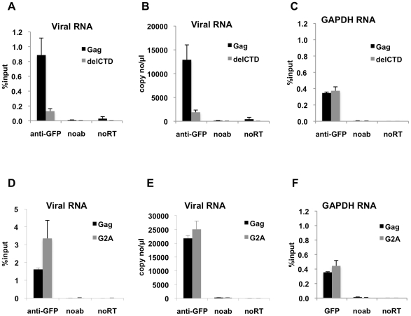
HIV-1 Gag drives a number of events during the genesis of virions and is the only viral protein required for the assembly of virus-like particles in vitro and in cells. Although a reasonable understanding of the processes that accompany the later stages of HIV-1 assembly has accrued, events that occur at the initiation of assembly are less well defined. In this regard, important uncertainties include where in the cell Gag first multimerizes and interacts with the viral RNA, and whether Gag-RNA interaction requires or induces Gag multimerization in a living cell. To address these questions, we developed assays in which protein crosslinking and RNA/protein co-immunoprecipitation were coupled with membrane flotation analyses in transfected or infected cells. We found that interaction between Gag and viral RNA occurred in the cytoplasm and was independent of the ability of Gag to localize to the plasma membrane. However, Gag:RNA binding was stabilized by the C-terminal domain (CTD) of capsid (CA), which participates in Gag-Gag interactions. We also found that Gag was present as monomers and low-order multimers (e.g. dimers) but did not form higher-order multimers in the cytoplasm. Rather, high-order multimers formed only at the plasma membrane and required the presence of a membrane-binding signal, but not a Gag domain (the CA-CTD) that is essential for complete particle assembly. Finally, sequential RNA-immunoprecipitation assays indicated that at least a fraction of Gag molecules can form multimers on viral genomes in the cytoplasm. Taken together, our results suggest that HIV-1 particle assembly is initiated by the interaction between Gag and viral RNA in the cytoplasm and that this initial Gag-RNA encounter involves Gag monomers or low order multimers. These interactions per se do not induce or require high-order Gag multimerization in the cytoplasm. Instead, membrane interactions are necessary for higher order Gag multimerization and subsequent particle assembly in cells.


Conflict of interest statement

The authors have declared that no competing interests exist.

Figures

Figure 1
Figure 1. Efficient immunoprecipitation of HIV-1 genomes by MS2-GFP or Gag-GFP.
(A and B) Lysates of 293T cells coexpressing V1B-MS2 RNA and either MS2-GFP (A) or Gag-GFP (B) were prepared at 24-h post-transfection. RNA-protein complexes were immunoprecipitated using anti-GFP antibodies. Immunoprecipitations without antibodies (noab) were carried in parallel. Immunoprecipitated V1B-MS2 RNA was quantitated by qRT-PCR and is represented as fraction of input RNA prior to immunoprecipitation (% input). (C) RNA-IP assay was performed as in (B) where the viral genome was either V1B or V1BΔΨ. (D) RNA-IP assay in 293T cells coexpressing V1B-MS2 viral RNA and Gag-GFP or untagged Gag. (E) RNA-IP assay was performed as in (B) but an immunoprecipitation using anti-FLAG antibodies was included as negative control. (F) 293T cells separately expressing Gag-GFP or V1B-MS2 viral RNA were mixed before cell lysis followed by immunoprecipitation (gray bars). RNA-IP assay was then performed as in (B). Immunoprecipitations from 293T cells that were cotransfected with Gag-GFP and V1B-MS2 plasmids were carried in parallel (black bars). Data in (A-F) represents the average of two independent experiments, where error bars indicate the range between the averages of two experiments. RNA extracted from 10% of the cell lysate that was used in immunoprecipitation was subjected to qRT-PCR without reverse transcription (noRT) to control for the presence of contaminating viral DNA.
Figure 2
Figure 2. Interaction of Gag with HIV-1 genome is enhanced by an intact CA domain but not by Gag myristoylation.
(A-C) V1B-MS2 viral RNA bound to either Gag-GFP (black bars) or Gag-delCTD-GFP (gray bars) was immunoprecipitated by anti-GFP antibodies from lysates of 293T cells. Immunoprecipitations without antibodies (noab) were included in parallel as controls. (D-F) V1B-MS2 viral RNA bound to either Gag-GFP (black bars) or Gag-G2A-GFP (gray bars) was immunoprecipitated by anti-GFP antibodies as in (A-C). Immunoprecipitated RNA was quantitated by qRT-PCR to detect viral RNA (A, B, D, E) or cellular GAPDH RNA (C, F) as described in Materials & Methods and is represented either as fraction of input material prior to immunoprecipitation (% input) or copies of DNA per µl of reverse transcribed template (copy no/µl). Data represents the average of two independent experiments, where error bars indicate the range between the averages of two experiments. noRT =  as explained in legend to Figure 1.
Figure 3
Figure 3. Immunoprecipitation of HIV genomic RNA from membrane and cytoplasmic fractions by Gag, G2A-Gag and Gag-delCTD.
(A-E) 293T cells coexpressing V1B-MS2 and either Gag-GFP, G2A-Gag-GFP or Gag-ΔCTD-GFP were processed for subcellular fractionation at 24-h post-transfection. Ten fractions from the top of the membrane flotation gradient were collected. Total RNA and proteins from these fractions were isolated as explained in Materials & Methods. (A and C) V1B-MS2 (A) and GAPDH (C) RNA in fractions were quantitated by qRT-PCR. Data is represented as the relative number of copies of cDNA with as compared to the tenth, cytoplasmic fraction of Gag-GFP sample that was arbitrarily set to a value of 100%. (B) Western blot analysis of Gag-GFP, G2A-Gag-GFP and Gag-ΔCTD-GFP in sucrose fractions and in post-nuclear supernatant (PNS) using anti-HIV-1 MA antibodies. (D and E) After subcellular fractionation, immunoprecipitations from the membrane (D) and cytoplasmic (E) fractions were performed using anti-GFP antibodies as explained in Materials & Methods. Parallel immunoprecipitations were carried in the absence of antibodies (noab). Immunoprecipitated V1B-MS2 RNA is quantitated by qRT-PCR and is represented as a fraction of input material prior to immunoprecipitation (% input, [D, E]). Data in (A, C, D, E) represents the average of two independent experiments where error bars indicate the range. noRT =  as explained in legend to Figure 1.
Figure 4
Figure 4. Immunoprecipitation of genomic viral RNA from membrane and cytoplasmic fractions of HIV-1 infected MT2 cells.
(A-F) MT2 cells were infected with VSV-G pseudotyped NL4-3 MA-YFP/PR- at MOI = 1 and processed for subcellular fractionation at 48-h post-infection. Ten fractions from the top of the membrane flotation gradient were collected. Total RNA and proteins from these fractions were isolated as explained in Materials & Methods. (A) Western blot analysis of MA-YFP Gag in sucrose fractions and in post-nuclear supernatant (PNS) using anti-HIV-1 CA antibodies. (B and C) Viral (B) and GAPDH (C) RNA in fractions was quantitated by qRT-PCR. Data is represented as the relative number of copies of cDNA as compared to the tenth, cytoplasmic fraction sample that was arbitrarily set to a value of 100%. (D-F) Immunoprecipitations from the membrane (black bars) and cytoplasmic (gray bars) fractions were performed using anti-GFP antibodies. Parallel immunoprecipitations without antibodies (noab) were included as controls. Immunoprecipitated viral RNA (D and E) or GAPDH RNA (F) was quantitated by qRT-PCR and is represented as either fraction of input material prior to immunoprecipitation (% input) or number of copies of cDNA per µl of PCR template. Data in (B-F) represents the average of two independent experiments where error bars indicate the range. noRT =  as explained in legend to Figure 1.
Figure 5
Figure 5. Multimerization of Gag, G2A-Gag, Gag-delCTD in plasma membrane and cytoplasm.
293T cells coexpressing FLAG and HA-tagged Gag or its derivatives (G2A-Gag or Gag-delCTD) together with V1B-MS2 viral RNA were cross-linked by treatment with 1mM EGS and used in membrane flotation assay. (A) Western blot analysis of Gag, G2A-Gag and Gag-delCTD in membrane fractions (lanes 1–3), cytoplasmic fractions (lanes 4–6), post-nuclear supernatants (PNS, lanes 7–9) and in mock crosslinked post-nuclear supernatants (lanes 10–12) using mouse anti-FLAG antibodies. (B) Quantitative analysis of membrane (lanes 1–3) and cytoplasmic (lanes 4–6) fractions of the western blot in (A). x-axis shows the pixel location and y-axis indicates the average pixel intensity in the lane that is analyzed. Degree of Gag multimerization is marked by subscripts (i.e. Gag3 corresponds to Gag trimers).
Figure 6
Figure 6. Gag molecules form multimers on viral RNA.
293T cells coexpressing FLAG and HA-tagged Gag or its derivatives (G2A-Gag or Gag-delCTD) together with V1B-MS2 viral RNA were used in seq-RNA-IP analysis after crosslinking with EGS and formaldehyde. (A, B) Viral RNA that was eluted after the first immunoprecipitation with anti-HA or anti-FLAG antibodies, or in the absence of antibodies (noab) is quantitated by qRT-PCR as in previous figures. (C, D) RNA-protein complexes obtained from the first immunoprecipitation (A, B) were used in a subsequent immunoprecipitation. Order of immunoprecipitations is indicated on the x-axis. Data is represented as either number of copies of cDNA per µl of PCR template (A, C) or fraction of input material (% input, B, D). Data in (A-D) shows the average of two independent experiments where error bars indicate the range.

Similar articles

Cited by

References

    1. Harrison GP, Lever AM. The human immunodeficiency virus type 1 packaging signal and major splice donor region have a conserved stable secondary structure. J Virol. 1992;66:4144–4153. - PMC - PubMed
    1. Lever A, Gottlinger H, Haseltine W, Sodroski J. Identification of a sequence required for efficient packaging of human immunodeficiency virus type 1 RNA into virions. J Virol. 1989;63:4085–4087. - PMC - PubMed
    1. Luban J, Goff SP. Mutational analysis of cis-acting packaging signals in human immunodeficiency virus type 1 RNA. J Virol. 1994;68:3784–3793. - PMC - PubMed
    1. Clever J, Sassetti C, Parslow TG. RNA secondary structure and binding sites for gag gene products in the 5′ packaging signal of human immunodeficiency virus type 1. J Virol. 1995;69:2101–2109. - PMC - PubMed
    1. Finzi A, Orthwein A, Mercier J, Cohen EA. Productive human immunodeficiency virus type 1 assembly takes place at the plasma membrane. J Virol. 2007;81:7476–7490. - PMC - PubMed

Publication types

MeSH terms