

Effects of photodynamic therapy on the endocytic pathway
- PMID: 21125114
- PMCID: PMC3069131
- DOI: 10.1039/c0pp00276c
Effects of photodynamic therapy on the endocytic pathway
Abstract
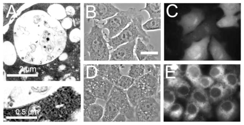
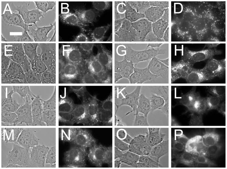
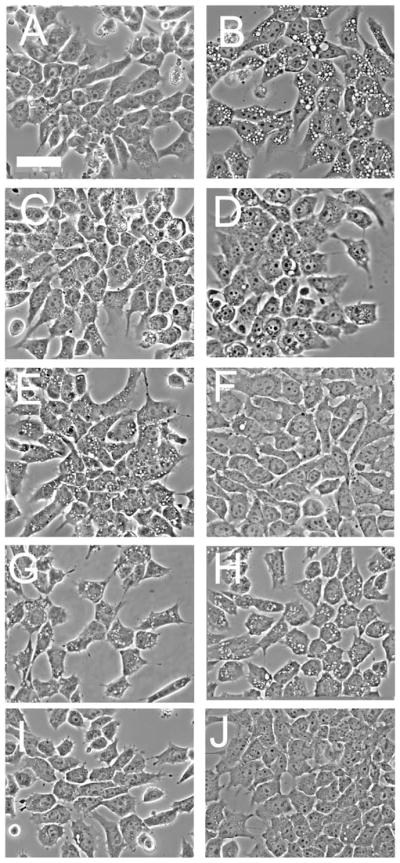
In this report, we describe an effect of photodynamic therapy (PDT) on membrane trafficking in murine 1c1c7 hepatoma cells. A brief exposure of 1c1c7 cells to a 20 nM concentration of the phosphatidylinositol kinase class-3 antagonist wortmannin led to the rapid appearance of cytoplasmic vacuoles. Fluorescence monitoring of plasma membrane-associated 1-[4-(trimethylamino)phenyl]-6-phenylhexa-1,3,5-triene (TDPH) over time demonstrated that the wortmannin-induced vacuoles were derived from endocytosed plasma membrane. Low-dose photodamage catalyzed by the lysosomal photosensitizer NPe6, prior to the addition of wortmannin, prevented formation of these vacuoles. NPe6 was found to suppress for several hours the normal trafficking of TDPH-labeled plasma membrane to the cytosol, and the formation of punctate TDPH-labeled cytoplasmic vesicles. The ability of NPe6-induced photodamage to suppress wortmannin-induced vacuolization occurred under conditions that did not disrupt lysosomes and were at or below the threshold of cytostatic/cytotoxic effects. Furthermore, the suppressive effects of NPe6-PDT were not prevented by inclusion of an agent that stabilized lysosomal membranes, or by E64d, an inhibitor of lysosomal cathepsin proteases. Mitochondrial photodamage was less effective at preventing wortmannin-induced vacuole formation and PDT directed against the ER had no effect. The role of photodamage to the endocytic pathway may be a hitherto unexplored effect on cells that selectively accumulate photosensitizing agents. These results indicate that photodamage directed against endosomes/lysosomes has effects independent of the release of lysosomal proteases.
Figures


Similar articles
-
Inhibition of endocytic processes by photodynamic therapy.Lasers Surg Med. 2011 Sep;43(7):542-7. doi: 10.1002/lsm.21067. Lasers Surg Med. 2011. PMID: 22057481 Free PMC article.
-
Effects of endosomal photodamage on membrane recycling and endocytosis.Photochem Photobiol. 2011 May-Jun;87(3):699-706. doi: 10.1111/j.1751-1097.2011.00890.x. Epub 2011 Feb 10. Photochem Photobiol. 2011. PMID: 21208213 Free PMC article.
-
ATG7 deficiency suppresses apoptosis and cell death induced by lysosomal photodamage.Autophagy. 2012 Sep;8(9):1333-41. doi: 10.4161/auto.20792. Epub 2012 Aug 14. Autophagy. 2012. PMID: 22889762 Free PMC article.
-
Photochemical internalisation: a novel drug delivery system.Tumour Biol. 2002 Mar-Apr;23(2):103-12. doi: 10.1159/000059713. Tumour Biol. 2002. PMID: 12065848 Review.
-
Apoptosis, Paraptosis and Autophagy: Death and Survival Pathways Associated with Photodynamic Therapy.Photochem Photobiol. 2019 Jan;95(1):119-125. doi: 10.1111/php.12952. Epub 2018 Jul 17. Photochem Photobiol. 2019. PMID: 29882356 Free PMC article. Review.
Cited by
-
Development of a new minimally invasive phototherapy for lung cancer using antibody-toxin conjugate.Thorac Cancer. 2023 Mar;14(7):645-653. doi: 10.1111/1759-7714.14776. Epub 2023 Jan 19. Thorac Cancer. 2023. PMID: 36655546 Free PMC article.
-
Enhanced efficacy of photodynamic therapy via a sequential targeting protocol.Photochem Photobiol. 2014 Jul-Aug;90(4):889-95. doi: 10.1111/php.12270. Epub 2014 Apr 1. Photochem Photobiol. 2014. PMID: 24617972 Free PMC article.
-
Lysosomal signaling enhances mitochondria-mediated photodynamic therapy in A431 cancer cells: role of iron.Photochem Photobiol. 2012 Mar-Apr;88(2):461-8. doi: 10.1111/j.1751-1097.2012.01081.x. Epub 2012 Jan 25. Photochem Photobiol. 2012. PMID: 22220628 Free PMC article.
-
Inhibition of endocytic processes by photodynamic therapy.Lasers Surg Med. 2011 Sep;43(7):542-7. doi: 10.1002/lsm.21067. Lasers Surg Med. 2011. PMID: 22057481 Free PMC article.
-
Effects of endosomal photodamage on membrane recycling and endocytosis.Photochem Photobiol. 2011 May-Jun;87(3):699-706. doi: 10.1111/j.1751-1097.2011.00890.x. Epub 2011 Feb 10. Photochem Photobiol. 2011. PMID: 21208213 Free PMC article.
References
-
- Oleinick NL, Morris RL, Belichenko I. The role of apoptosis in response to photodynamic therapy: what, where, why, and how. Photochem Photobiol Sci. 2002;1:1–21. - PubMed
-
- Reaves BJ, Bright NA, Mullock BM, Luzio JP. The effect of wortmannin on the localisation of lysosomal type I integral membrane glycoproteins suggests a role for phosphoinositide 3-kinase activity in regulating membrane traffic late in the endocytic pathway. J Cell Sci. 1996;109:749–762. - PubMed
Publication types
MeSH terms
Substances
Grants and funding
LinkOut - more resources
Full Text Sources