. 2011 Mar;349(1-2):125-37.
doi: 10.1007/s11010-010-0667-3. Epub 2010 Dec 2.

Treatment of cultured human astrocytes and vascular endothelial cells with protein kinase CK2 inhibitors induces early changes in cell shape and cytoskeleton

Affiliations

Treatment of cultured human astrocytes and vascular endothelial cells with protein kinase CK2 inhibitors induces early changes in cell shape and cytoskeleton

A A Kramerov et al. Mol Cell Biochem. 2011 Mar.

Abstract

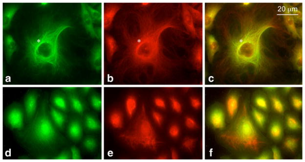
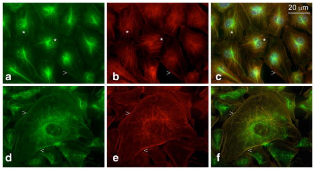
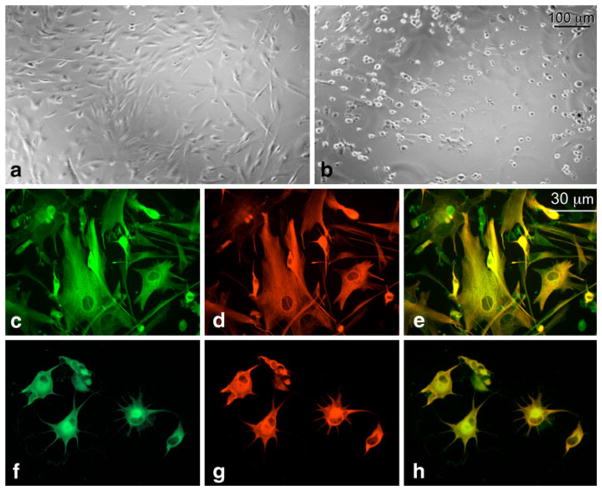
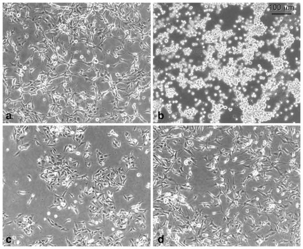
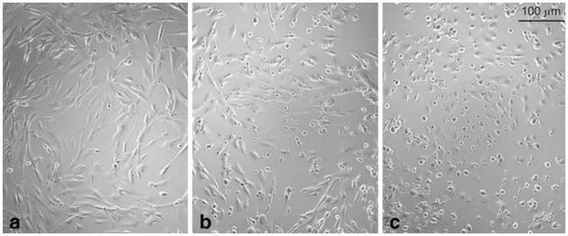
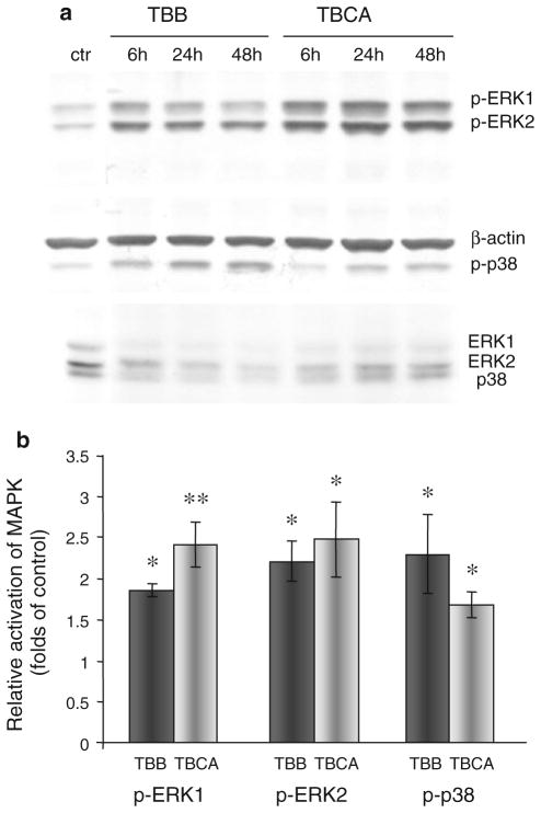
Ubiquitous protein kinase CK2 is a key regulator of cell migration, proliferation and tumor growth. CK2 is abundant in retinal astrocytes, and its inhibition suppresses retinal neovascularization in a mouse retinopathy model. In human astrocytes, CK2 co-distributes with GFAP-containing intermediate filaments, which implies its association with cytoskeleton. Contrary to astrocytes, CK2 is co-localized in microvascular endothelial cells (HBMVEC) with microtubules and actin stress fibers, but not with vimentin-containing intermediate filaments. Specific CK2 inhibitors (TBB, TBI, TBCA and DMAT) and nine novel CK2 inhibiting compounds (TID43, TID46, Quinolone-7, Quinolone-39, FNH28, FNH62, FNH64, FNH68 and FNH74) were tested at 10-200 μM for their ability to induce morphological alterations in cultured human astrocytes (HAST-40), and HBMVEC (For explanation of the inhibitor names, see "Methods" section). CK2 inhibitors caused dramatic changes in shape of cultured cells with effective inhibitor concentrations between 50 and 100 μM. Attached cells retracted, acquired shortened processes, and eventually rounded up and detached. CK2 inhibitor-induced morphological alterations were completely reversible and were not blocked by caspase inhibition. However, longer treatment or higher inhibitor concentration did cause apoptosis. The speed and potency of the CK2 inhibitors effects on cell shape and adhesion were inversely correlated with serum concentration. Western analyses showed that TBB and TBCA elicited a significant (about twofold) increase in the activation of p38 and ERK1/2 MAP kinases that may be involved in cytoskeleton regulation. This novel early biological cell response to CK2 inhibition may underlie the anti-angiogenic effect of CK2 suppression in the retina.


Figures

Fig. 1
Fig. 1
CK2 association with the cytoskeleton in HBMVEC as revealed by double immunostaining with anti-CK2 (green) and anti-tubulin (a–c) or anti-vimentin (d–f) antibodies (both red). CK2 co-distributed with the tubulin-containing cytoskeleton (especially in the perinuclear region, marked by the asterisk), but not with cytoskeletal elements that contained vimentin. (Color figure online)
Fig. 2
Fig. 2
Co-localization of CK2 and F-actin in HBMVEC by double staining with anti-CK2 (green) and rhodamine-phalloidin (red). Yellow color (in c and f) demonstrates co-distribution of CK2 with actin microfilaments of stress fibers (a–c, marked by the asterisks) and cortical actin ring (d–f, marked by arrowheads). The results are representative of three experiments. (Color figure online)
Fig. 3
Fig. 3
Dramatic cell shape changes induced by CK2 inhibitor TBB in HAST-40 cells: a control cells, treated with 0.05% DMSO, b cells treated with 100 μM TBB. The results are representative of five independent experiments. Co-distribution of CK2 (green color) and GFAP (red color) is preserved in HAST-40 cells treated with 0.1 mM TBB. c–e Control DMSO-treated cells, f–h TBB-treated cells. The results are representative of three experiments. (Color figure online)
Fig. 4
Fig. 4
The reversal of cell shape changes induced after CK2 inhibition by TBB. The cultured U87MG cells were treated with DMSO (a), or 0.1 mM TBB (b) for 18 h, followed by incubation in medium without TBB for 6 h (c) or 24 h (d). After 6 h of incubation in the medium without TBB, formerly round cells (b) acquired nearly normal polygonal morphology (c) although they remained less spread than the cells after 24-h recovery period (d). The results are representative of three independent experiments
Fig. 5
Fig. 5
Partial serum deprivation facilitates the development of TBB-induced cell shape changes in HAST-40 cultured cells. Cells were incubated with medium containing 0.5% FBS and DMSO (a), or treated with 0.1 mM TBB in the presence of 5% (b) or 0.5% (c) FBS for 6 h. Virtually all the cells treated with TBB in the presence of 0.5% FBS retracted and turned round, whereas the majority of the cells treated at 5% FBS failed to do so. The results are representative of four experiments
Fig. 6
Fig. 6
MAPK activation after CK2 inhibition. a Western analyses of activated signaling molecules ERK1, ERK2, and p38 MAPK after treatment of HBMVEC with CK2 inhibitors TBB and TBCA for 6–48 h. Phosphospecific p38 or ERK1 and ERK2 antibodies were used to visualize phosphorylated p38 MAPK and ERKs. Antibodies to β-actin or unphosphorylated ERKs and p38 were used to ensure equal loading. The typical blot is representative of three independent experiments. ctr, DMSO-treated control. b Quantitation of CK2 inhibitor-induced phosphorylation of MAPK. The bar graph represents average ± SEM of pooled values (n = 5) of densitometric scans. *P < 0.05, **P < 0.01 compared with control values (taken as 1) by paired two-tailed t test

References

    1. Meggio F, Pinna LA. One-thousand-and-one substrates of protein kinase CK2? FASEB J. 2003;17:349–368. - PubMed
    1. Guerra B, Issinger O-G. Protein kinase CK2 in human disease. Curr Med Chem. 2008;15:1870–1886. - PubMed
    1. Trembley JH, Wang G, Unger G, Slaton J, Ahmed K. Protein kinase CK2 in health and disease: CK2: a key player in cancer biology. Cell Mol Life Sci. 2009;66:186–1858. - PMC - PubMed
    1. Ljubimov AV, Caballero S, Aoki A, Pinna LA, Grant MB, Castellon R. Involvement of protein kinase CK2 in angi-ogenesis and retinal neovascularization. Invest Ophthalmol Vis Sci. 2004;45:4583–4591. - PMC - PubMed
    1. Kramerov AA, Saghizadeh M, Pan H, Kabosova A, Montenarh M, Ahmed K, Penn JS, Chan CK, Hinton DR, Grant MB, Ljubimov AV. Expression of protein kinase CK2 in astroglial cells of normal and neovascularized retina. Am J Pathol. 2006;168:1722–1736. - PMC - PubMed

Publication types

MeSH terms

LinkOut - more resources