Review
. 2011 Oct 1;15(7):2011-35.
doi: 10.1089/ars.2010.3603. Epub 2011 Apr 21.

Antioxidants, redox signaling, and pathophysiology in schizophrenia: an integrative view

Affiliations
Review

Antioxidants, redox signaling, and pathophysiology in schizophrenia: an integrative view

Jeffrey K Yao et al. Antioxid Redox Signal. .

Abstract

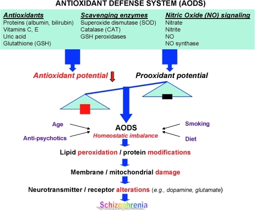
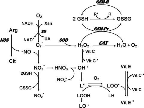
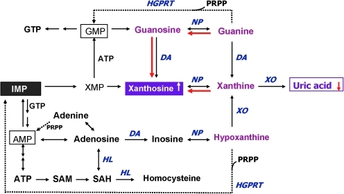
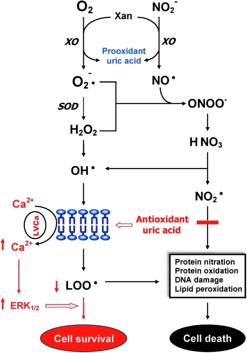
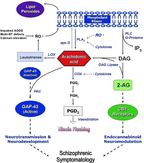
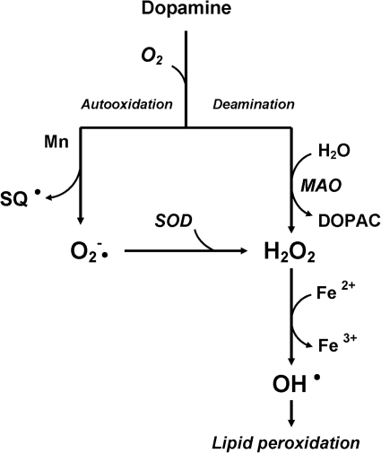
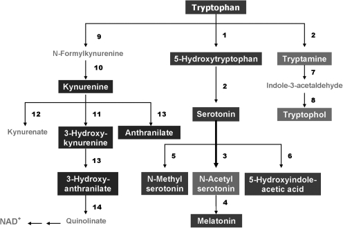
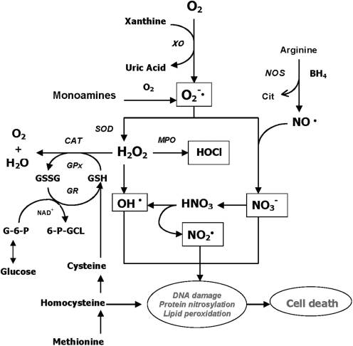
Schizophrenia (SZ) is a brain disorder that has been intensively studied for over a century; yet, its etiology and multifactorial pathophysiology remain a puzzle. However, significant advances have been made in identifying numerous abnormalities in key biochemical systems. One among these is the antioxidant defense system (AODS) and redox signaling. This review summarizes the findings to date in human studies. The evidence can be broadly clustered into three major themes: perturbations in AODS, relationships between AODS alterations and other systems (i.e., membrane structure, immune function, and neurotransmission), and clinical implications. These domains of AODS have been examined in samples from both the central nervous system and peripheral tissues. Findings in patients with SZ include decreased nonenzymatic antioxidants, increased lipid peroxides and nitric oxides, and homeostatic imbalance of purine catabolism. Reductions of plasma antioxidant capacity are seen in patients with chronic illness as well as early in the course of SZ. Notably, these data indicate that many AODS alterations are independent of treatment effects. Moreover, there is burgeoning evidence indicating a link among oxidative stress, membrane defects, immune dysfunction, and multineurotransmitter pathologies in SZ. Finally, the body of evidence reviewed herein provides a theoretical rationale for the development of novel treatment approaches.


Figures

FIG. 1.
FIG. 1.
A schematic diagram linking homeostatic imbalance in antioxidant defense system to pathophysiology in schizophrenia (SZ). (To see this illustration in color the reader is referred to the web version of this article at www.liebertonline.com/ars).
FIG. 2.
FIG. 2.
Possible mechanisms involving production and removal of oxygen and nitrogen free radicals in mammalian cells. Molecular oxygen can be converted to superoxide radicals (O2•) in the presence of xanthine oxidase (XO). Subsequently, superoxide dismutase (SOD) catalyzes the conversion of superoxide radicals to hydrogen peroxide (H2O2). Catalase (CAT) and glutathione peroxidase (GSH-Px) convert hydrogen peroxide to water. Glutathione (GSH) is utilized by GSH-Px to yield the oxidized form of glutathione (GSSG), which is converted back to GSH by glutathione reductase (GR). Hydrogen peroxide is susceptible to autoxidation to form hydroxyl radicals (OH•), particularly in the presence of metal catalysts such as iron. In addition, nitric oxide (NO), which is the product of a five-electron oxidation of the amino acid L-arginine, can also produce hydroxyl radicals as well as nitrogen dioxide radical. On the other hand, α-tocopherol (vitamin E) has the ability to inhibit lipid peroxidation as a chain-breaking antioxidant. Vitamin E radicals can be recycled back to their native form by ascorbic acid (vitamin C).
FIG. 3.
FIG. 3.
Altered purine catabolism in first-episode neuroleptic-naïve patients with SZ. Red arrows indicate shifts toward an increase of xanthosine and a decrease of uric acid productions in first-episode neuroleptic-naïve patients with SZ patients at baseline. Reactions shown with dotted lines represent the salvage pathways, which purine bases can be reutilized resulting in considerably energy saving for the cell. Reprinted with permission from Yao et al. (284). (To see this illustration in color the reader is referred to the web version of this article at www.liebertonline.com/ars).
FIG. 4.
FIG. 4.
Dual role of uric acid in the antioxidant defense system. Uric acid can neutralize peroxynitrite and hydroxyl radicals to inhibit protein nitration and lipid peroxidation, respectively. At increased levels, however, uric acid may be considered as a marker of oxidative stress due to accumulation of reactive oxygen species. Reprinted with permission from Yao et al. (284). (To see this illustration in color the reader is referred to the web version of this article at www.liebertonline.com/ars).
FIG. 5.
FIG. 5.
Correlations of GSH-Px to glutathione reductase (GSH-R) activities in postmortem caudate region. (A) Control subjects without mental disorders, (B) control subjects with bipolar and/or depression, and (C) patients with SZ. Reprinted with permission from Yao et al. (286).
FIG. 6.
FIG. 6.
Correlations of GSSG or GSH-R activities to age in postmortem caudate region. (A) Control subjects without mental disorders, and (B) patients with SZ. Reprinted with permission from Yao et al. (286).
FIG. 7.
FIG. 7.
Schematic membrane structure illustrating the key components and their functional role in cell–cell interaction, interaction with environmental factors, and receptor-mediated signal transduction. Membrane model shows the phospholipid bilayer with embedded in it the receptors for neurotransmitters, physiological mediators and growth factors, transporters for ions and nutritional ingredients, and signal transduction machinery. Phospholipid bilayer is made up of four major phospholipids, phosphatidylcholine (PC), phosphatidylethanolamine (PE), phosphatidylinositol (PI), and phosphatidylserine (PS), which are asymmetrically localized, that is, PC predominantly on outer lipid layer and PE, PI, and PS on inner lipid layer. PE, PI, and PS are highly enriched in arachidonic acid (AA) and docosahexaenoic acid (DHA), which are released by receptor-mediated phospholipases (PLAs). AA and DHA, and their metabolic products, diacylglycerol (DAG), inositol polyphosphates (IPPs), and prostaglandins work as second messengers and physiological mediators, including gene modulation, and thus lead to adaptive and maladaptive cellular changes. Reprinted with permission from Mahadik and Yao (151).
FIG. 8.
FIG. 8.
A typical in vivo 31P magnetic resonance spectrum from the prefrontal region of a healthy subject. The left panel shows the voxel placement for the spectral acquisition, and the right panel shows the spectrum. The x axis represents the frequency in parts per million (ppm). ATP, adenosine triphosphate; PCr, phosphocreatine; PDE, phosphodiester; Pi, inorganic phosphate; PME, phosphomonoesters. (To see this illustration in color the reader is referred to the web version of this article at www.liebertonline.com/ars).
FIG. 9.
FIG. 9.
A putative model integrating lipid peroxidation, phospholipids turnover, AA signaling, and SZ symptomatology. As shown, several possible mechanisms can lead to increased phospholipid breakdown and AA release, including decreased AA incorporation and increased phospholipase activities (PLA2 and PLC), possibly resulting from increased oxidative stress and cytokine release. The resulting changes in AA level could then affect more downstream processes, including neurodevelopment via growth-associated protein (GAP)-43, neurotransmitter homeostasis, phosphatidylinositol signaling, and neuromodulatory actions of endocannabinoids. It is proposed that the specific behavioral symptomatology of SZ is related mostly to the effect of AA changes on the neurochemistry of deaminase, glutamate release, and circulating levels of the endocannabinoids anandamide and 2-arachidonoylglycerol (2-AG). In addition, alterations in AA may also affect the inflammatory response, which can then affect PLA2 release via cytokines, further exacerbating phospholipid turnover and AA release. Hence, in the current conceptualization, AA is at a nexus point in the cascade leading to the syndrome of SZ, and represents a common biochemical pathway leading to the highly heterogeneous symptomatology of psychosis. Reprinted with permission from Skosnik and Yao (236). (To see this illustration in color the reader is referred to the web version of this article at www.liebertonline.com/ars).
FIG. 10.
FIG. 10.
Autooxidation and oxidative deamination of dopamine. In the presence of Mn, the auto-oxidation of dopamine produces semiquinones (SQ) and superoxide radicals (O2•), as well as H2O2, which is readily converted to OH• in the presence of Fe2+. On the other hand, the enzymatic oxidation of dopamine by monoamine oxidase (MAO) can also produce H2O2 and, subsequently, generate the toxic OH•.
FIG. 11.
FIG. 11.
Tryptophan metabolic pathways. The bolded arrow indicates that pathway may be upregulated in first-episode neuroleptic-naïve patients with SZ. 1, tryptophan hydroxylase; 2, aromatic L-amino acid decarboxylase; 3, serotonin N-acetyltransferase; 4, 5-hydroxyindole-O-methyltransferase; 5, serotonin N-methyltransferase; 6, monoamine oxidase and aldehyde dehydrogenase; 7, monoamine oxidase; 8, alcohol dehydrogenase; 9, tryptophan 2,3-dioxygenase; 10, formamidase; 11, kynurenine 3-monooxygenase; 12, kynurenine transaminase; 13, kynureninase; 14, 3-hydroxyanthranilate oxigenase. Reprinted with permission from Yao et al. (283).
FIG. 12.
FIG. 12.
Antioxidant defense system involving multiple biochemical pathways. It is surmised that some, or all, of these alterations in purine catabolism, neurotransmission, glutathione redox coupling, glucose phosphorylation, one-carbon metabolism, and nitric oxide synthase activation may contribute to oxidative stress and membrane dysfunction in SZ. Reprinted with permission from Yao and Reddy (281).
FIG. 13.
FIG. 13.
Separation of low-molecular-weight, redox-active compounds by high-pressure liquid chromatography coupled with a Coulometric Multielectrode Array System. (A) Flow rate and mobile phase gradient profile; (B) 16-channel chromatograms obtained by separation of standard mixture in a single column (ESA Meta-250, 5 μm ODS, 250 × 4.6 mm ID) under a 150-min gradient elution that ranged from 0% to 20% MPB with a fixed flow rate of 0.5 ml/min (A). The temperature of both cells and column was maintained at 25°C. The Coulometric Multielectrode Array System was set to have increments from 0 to 900 mV in 60 mV steps. Reprinted with permission from Yao and Reddy (281).

References

    1. Abdalla DS. Manteiro HP. Olivera JA. Bechara EJ. Activities of superoxide dismutase and glutathione peroxidase in schizophrenic and manic depressive patients. Clin Chem. 1986;32:805–807. - PubMed
    1. Akyol O. Zoroglu S. Armutcu F. Sahin S. Gurel A. Nitric oxide as a physiopathological factor in neuropsychiatric disorders. In Vivo. 2004;18:377–390. - PubMed
    1. Al-Chalabi BM. Thanoon IA. Ahmed FA. Potential effect of olanzapine on total antioxidant status and lipid peroxidation in schizophrenic patients. Neuropsychobiology. 2009;59:8–11. - PubMed
    1. Amminger GP. Schäfer MR. Papageorgiou K. Klier CM. Cotton SM. Harrigan SM. Mackinnon A. McGorry PD. Berger GE. Long-chain omega-3 fatty acids for indicated prevention of psychotic disorders: a randomized, placebo-controlled trial. Arch Gen Psychiatry. 2010;67:146–154. - PubMed
    1. Anderson ME. Glutathione: an overview of biosynthesis and modulation. Chem Biol Interact. 1998;111–112:1–14. - PubMed

Publication types

MeSH terms