. 2011 Sep 15;15(6):1507-15.
doi: 10.1089/ars.2010.3631. Epub 2011 Apr 8.

Adventitia-derived hydrogen peroxide impairs relaxation of the rat carotid artery via smooth muscle cell p38 mitogen-activated protein kinase

Affiliations

Adventitia-derived hydrogen peroxide impairs relaxation of the rat carotid artery via smooth muscle cell p38 mitogen-activated protein kinase

Thomas Cascino et al. Antioxid Redox Signal. .

Abstract

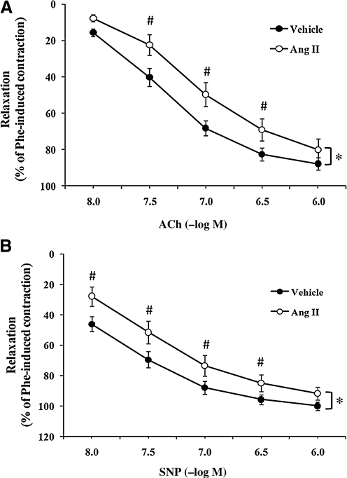
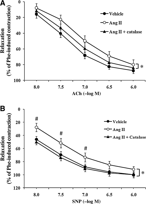
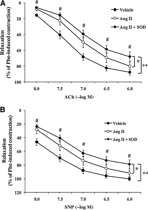
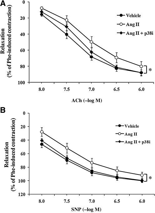
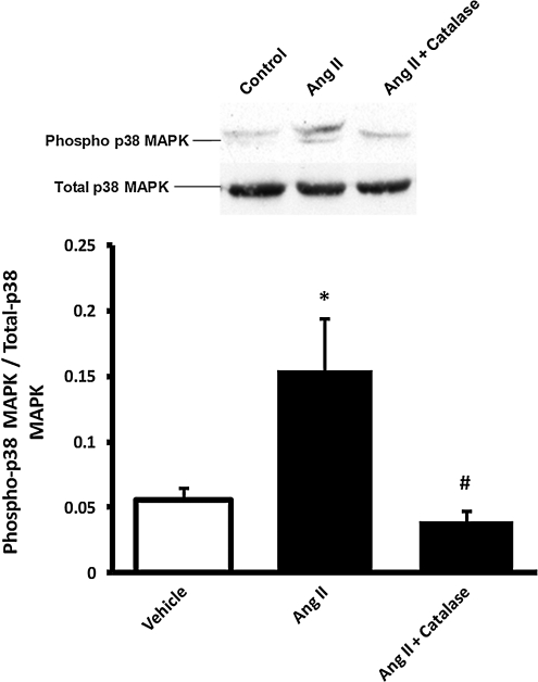
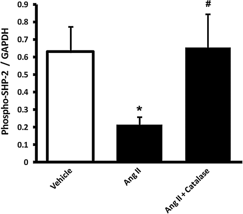
The role of adventitia-derived reactive oxygen species (ROS) in vascular disease and impaired vascular relaxation is not clear. Based on robust adventitial ROS generation and effects on MAPK involvement in vascular dysfunction, we hypothesized that adventitia-derived ROS hydrogen peroxide (H(2)O(2)) impairs vascular relaxation through activation of medial smooth muscle p38 MAPK. By using a novel in vivo model, the adventitial surface of rat carotid arteries was bathed in situ for 90 min with vehicle, angiotensin II (AngII; 500 nM), AngII+H(2)O(2)-scavenger catalase (3,000 U/ml), AngII+p38 MAPK inhibitor SB203580 (10 μM), or AngII+superoxide dismutase (SOD; 150 U/ml). After these in vivo treatments, ex vivo tone measurements on isolated vessels revealed that periadventitial application of AngII impaired both acetylcholine-induced (endothelium-dependent) and sodium nitroprusside-induced (endothelium-independent) relaxations. In vivo coincubation with catalase or SB203580 significantly improved, but SOD exacerbated AngII-induced impairment of in vitro endothelium-dependent and -independent vascular relaxations. Western blots of vascular media, separated from the adventitia, demonstrated increased medial p38 MAPK activation and decreased medial phosphatase SHP-2 activity in AngII-treated vessels. These effects were reversed by in vivo periadventitial addition of catalase. These findings provide the first evidence that adventitia-derived H(2)O(2) participates in vascular dysfunction through p38 MAPK activation and SHP-2 inhibition.


Figures

FIG. 1.
FIG. 1.
Periadventitial angiotensin II (AngII) treatment induced impairment of endothelium-dependent and endothelium-independent carotid artery relaxation. The perivascular layer of rat carotid arteries was incubated in situ for 90 min with vehicle (0.9% sodium chloride solution; n = 15) or AngII (500 nM; n = 7). Isolated carotid arteries were preconstricted with phenylephrine (Phe, 10−6 M). (A) Concentration–response curves for acetylcholine (Ach)-induced endothelium-dependent relaxation of isolated carotid arteries. (B) Concentration–response curves for sodium nitroprusside (SNP)-induced endothelium-independent relaxation of isolated carotid arteries. Data represent mean ± SEM. *p < 0.05 indicates significant differences in relaxation between vehicle and AngII groups. #p < 0.05 indicates significant differences at individual concentrations between vehicle and AngII.
FIG. 2.
FIG. 2.
Perivascular incubation of catalase improved AngII-induced impairment of vascular relaxation. The perivascular layer of rat carotid arteries was incubated in situ for 90 min with vehicle (n = 15), AngII (500 nM; n = 7) or AngII + catalase (3,000 U/ml; n = 6). Isolated carotid arteries were preconstricted with phenylephrine (10−6 M). (A) Concentration–response curves for ACh-induced endothelium-dependent relaxation of isolated carotid arteries. (B) Concentration–response curves for SNP-induced endothelium-independent relaxation of isolated carotid arteries. Data represent mean ± SEM. *p < 0.05 indicates significant differences in relaxation between AngII and AngII + catalase groups. #p < 0.05 indicates significant differences at individual concentrations between AngII and AngII + catalase.
FIG. 3.
FIG. 3.
Superoxide dismutase (SOD) did not improve AngII-induced endothelium-dependent and endothelium-independent relaxation. The perivascular layer of rat carotid arteries was incubated in situ for 90 min with vehicle (n = 15), AngII (500 nM; n = 7) or AngII + SOD (150 U/ml; n = 6). Isolated carotid arteries were preconstricted with phenylephrine (10−6 M). (A) Concentration–response curves for ACh-induced endothelium-dependent relaxation of isolated carotid arteries. (B) Concentration–response curves for SNP-induced endothelium-independent relaxation in isolated carotid arteries. Data represent mean ± SEM. *p < 0.05 indicates a significant difference in relaxation between AngII and AngII + SOD groups. **p < 0.05 indicates a significant difference in relaxation between vehicle and AngII + SOD groups. #p < 0.05 indicates significant differences at individual concentrations between vehicle and AngII + SOD.
FIG. 4.
FIG. 4.
The p38 MAPK inhibitor SB203580 improved AngII-induced impairment of endothelium-dependent and -independent relaxation. The perivascular layer of rat carotid arteries was incubated in situ for 90 min with vehicle (n = 15), AngII (500 nM; n = 7), or AngII + SB203580 (p38i, 10 μM; n = 9). Isolated carotid arteries were preconstricted with phenylephrine (10−6 M). (A) Concentration–response curves for ACh-induced endothelium-dependent relaxation in isolated carotid arteries. (B) Concentration–response curves for SNP-induced endothelium-independent relaxation in isolated carotid arteries. Data represent mean ± SEM. *p < 0.05 indicates significant differences in relaxation between AngII and AngII + p38i groups.
FIG. 5.
FIG. 5.
Perivascular AngII treatment induced medial p38 MAPK phosphorylation that was inhibited by co-treatment with catalase. The perivascular layer of rat carotid arteries was incubated in situ for 90 min with vehicle (n = 4), AngII (500 nM; n = 4), or AngII + catalase (3,000 U/ml; n = 4). Medial homogenates were subjected to Western blot with phospho- or total p38 MAPK antibody. Bar graphs represent averaged optical-density data expressed as a ratio of phospho- to total p38 MAPK. *p < 0.05 indicates a significant difference between vehicle and AngII groups. #p < 0.05 indicates a significant difference between AngII and AngII + catalase.
FIG. 6.
FIG. 6.
Perivascular AngII treatment induced medial SHP-2 dephosphorylation that was reversed by co-treatment with catalase. The perivascular layer of rat carotid arteries was incubated in situ for 90 min with vehicle (n = 4), AngII (500 nM; n = 4), or AngII + catalase (3,000 U/ml; n = 4). Medial homogenates were subjected to Western blot with phospho-SHP-2 (SHP-2-[Tyr542]) or GAPDH antibody. Bar graphs represent averaged optical-density data expressed as a ratio of phospho-SHP2 to GAPDH. *p < 0.05 indicates a significant difference between vehicle and AngII groups. #p < 0.05 indicates a significant difference between AngII and AngII + catalase.

References

    1. Ardanaz N. Beierwaltes WH. Pagano PJ. Comparison of H2O2-induced vasoconstriction in the abdominal aorta and mesenteric artery of the mouse. Vascul Pharmacol. 2007;47:288–294. - PubMed
    1. Ardanaz N. Beierwaltes WH. Pagano PJ. Distinct hydrogen peroxide-induced constriction in multiple mouse arteries: potential influence of vascular polarization. Pharmacol Rep. 2008;60:61–67. - PubMed
    1. Ardanaz N. Pagano PJ. Hydrogen peroxide as a paracrine vascular mediator: regulation and signaling leading to dysfunction. Exp Biol Med. 2006;231:237–251. - PubMed
    1. Azuma Y. Kosaka K. Kashimata M. Phospholipase D-dependent and -independent p38MAPK activation pathways are required for superoxide production and chemotactic induction, respectively, in rat neutrophils stimulated by fMLP. Eur J Pharmacol. 2007;568:260–268. - PubMed
    1. Azumi H. Inoue N. Takeshita S. Rikitake Y. Kawashima S. Hayashi Y. Itoh H. Yokoyama M. Expression of NADH/NADPH oxidase p22phox in human coronary arteries. Circulation. 1999;100:1494–1498. - PubMed

Publication types

MeSH terms

LinkOut - more resources