. 2011 May 15;14(10):1919-28.
doi: 10.1089/ars.2010.3768. Epub 2011 Mar 6.

Mitophagy selectively degrades individual damaged mitochondria after photoirradiation

Affiliations

Mitophagy selectively degrades individual damaged mitochondria after photoirradiation

Insil Kim et al. Antioxid Redox Signal. .

Abstract

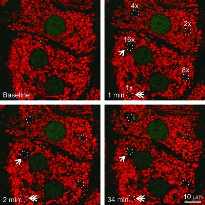
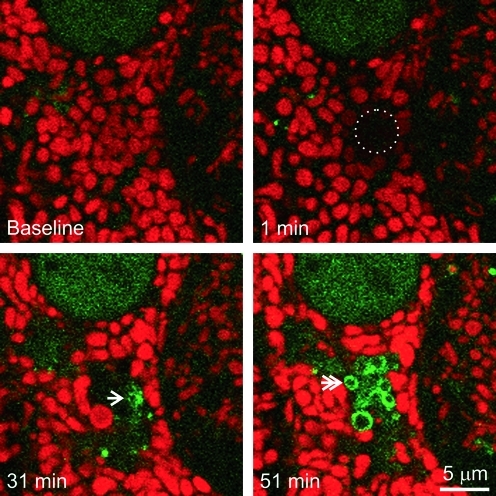
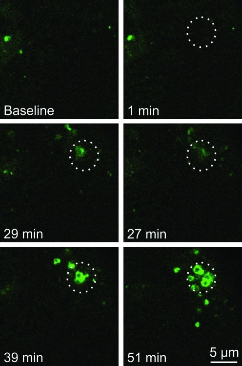
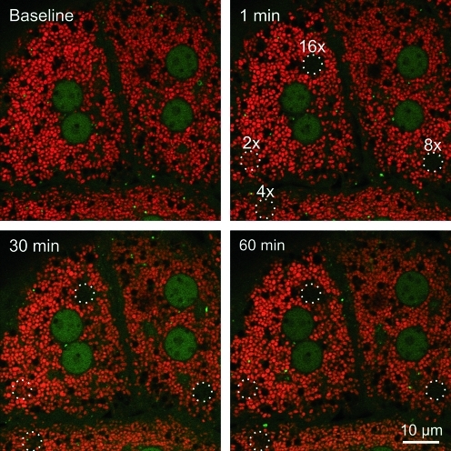
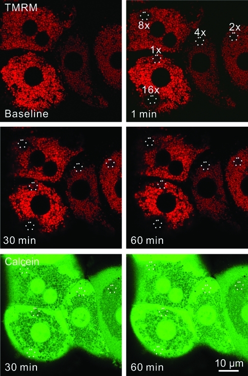
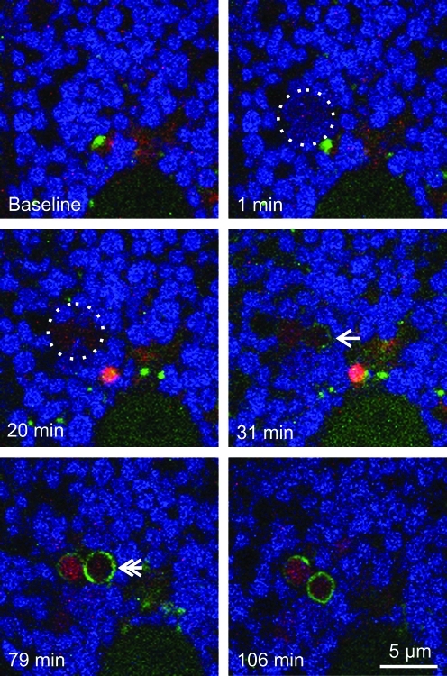
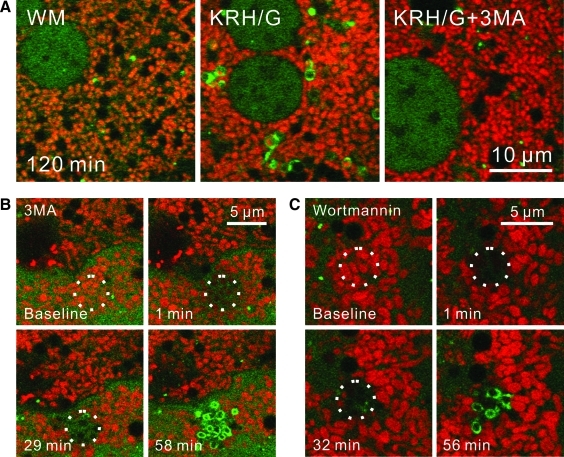
Damaged and dysfunctional mitochondria are proposed to be removed by autophagy. However, selective degradation of damaged mitochondria by autophagy (mitophagy) has yet to be experimentally verified. In this study, we investigated the cellular fate of individual mitochondria damaged by photoirradiation in hepatocytes isolated from transgenic mice expressing green fluorescent protein fused to microtubule-associated protein 1 light chain 3, a marker of forming and newly formed autophagosomes. Photoirradiation with 488-nm light induced mitochondrial depolarization (release of tetramethylrhodamine methylester [TMRM]) in a dose-dependent fashion. At lower doses of light, mitochondria depolarized transiently with re-polarization within 3 min. With greater light, mitochondrial depolarization became irreversible. Irreversible, but not reversible, photodamage induced autophagosome formation after 32±5 min. Photodamage-induced mitophagy was independent of TMRM, as photodamage also induced mitophagy in the absence of TMRM. Photoirradiation with 543-nm light did not induce mitophagy. As revealed by uptake of LysoTracker Red, mitochondria weakly acidified after photodamage before a much stronger acidification after autophagosome formation. Photodamage-induced mitophagy was not blocked by phosphatidylinositol 3-kinase inhibition with 3-methyladenine (10 mM) or wortmannin (100 nM). In conclusion, individual damaged mitochondria become selectively degraded by mitophagy, but photodamage-induced mitophagic sequestration occurs independently of the phosphatidylinositol 3-kinase signaling pathway, the classical upstream signaling pathway of nutrient deprivation-induced autophagy.


Figures

FIG. 1.
FIG. 1.
Mitochondrial damage occurs in a dose-dependent manner after exposure to 488-nm light. Hepatocytes isolated from GFP-LC3 transgenic mice were loaded with 200 nM TMRM for 30 min. Regions of the TMRM (red)-loaded hepatocytes were exposed to different amounts of 488-nm light at 100% power at 2.7, 5.5, 11, 22, and 44×104 times greater than the pixel illumination used for imaging. These illumination powers were labeled in ascending order as 1×, 2×, 4×, 8×, and 16×. The circles represent areas of photoirradiation. Images of red TMRM and green GFP-LC3 were collected before (baseline) and every minute after photoirradiation. Loss of TMRM fluorescence indicated depolarization of mitochondria. The double arrow indicates transient TMRM loss after photodamage at 1×. The arrow illustrates TMRM loss from the mitochondria just outside the zone of illumination at 16×. GFP-LC3, green fluorescence protein fused to microtubule-associated protein1 light chain 3; TMRM, tetramethyl rhodamine methyl ester. (To see this illustration in color the reader is referred to the web version of this article at www.liebertonline.com/ars).
FIG. 2.
FIG. 2.
Photodamaged mitochondria are degraded by mitophagy. A TMRM-loaded GFP-LC-3 hepatocyte was exposed to 16×illumination, as described in Figure 1. The circle indicates the area of photoirradiation. The arrow indicates first localization of GFP-LC3. The double arrow illustrates GFP-LC3 ring formation. (To see this illustration in color the reader is referred to the web version of this article at www.liebertonline.com/ars).
FIG. 3.
FIG. 3.
Photodamage-induced mitophagy is independent of TMRM. GFP-LC3 hepatocytes were exposed to 488-nm light at 16×relative power. The circles indicate the area of photoirradiation. (To see this illustration in color the reader is referred to the web version of this article at www.liebertonline.com/ars).
FIG. 4.
FIG. 4.
Exposure to 543-nm light does not induce mitophagy. TMRM (red)-loaded GFP-LC3 hepatocytes were exposed to different amounts of 543-nm light at 100% power–5.5, 11, 22, and 44×104 times higher than the power used for imaging. These pixel powers were labeled in ascending order as 2×, 4×, 8×, and 16×. The circles represent the areas of irradiation. Images of red TMRM and green GFP-LC3 were collected every minute before (baseline) and after photodamage. (To see this illustration in color the reader is referred to the web version of this article at www.liebertonline.com/ars).
FIG. 5.
FIG. 5.
Inner membrane permeabilization occurs after photodamage. Hepatocytes from a wild-type mouse were loaded with TMRM and exposed to 488-nm laser light at relative powers of 1×, 2×, 4×, 8×, and 16×, as described in Figure 1. Images were collected before (baseline) and after photoirradiation. After exposure, hepatocytes were loaded with 1 μM calcein acetoxymethyl ester for 10 min, and images of red TMRM and green calcein fluorescence were collected. The circles indicate areas of photoirradiation. (To see this illustration in color the reader is referred to the web version of this article at www.liebertonline.com/ars).
FIG. 6.
FIG. 6.
Mitophagosomes formed after photodamage acidify. GFP-LC3 hepatocytes were loaded with 300 nM MFFR and 500 nM LTR for 30 min. Circles show the area of photoirradiation with 488-nm light at a relative intensity of 16 ×. After photoirradiation, mitochondria lost MFFR fluorescence (pseudocolored blue) and took up red LTR fluorescence, which signified acidication. Arrow indicates localization of green GFP-LC3 to damaged mitochondria. Double arrow indicates GFP-LC3 rings (autophagosomes) around LTR-labeled photodamaged mitochondria. LTR, Lysotracker Red. (To see this illustration in color the reader is referred to the web version of this article at www.liebertonline.com/ars).
FIG. 7.
FIG. 7.
3-Methyladenine and wortmanin do not prevent mitophagy after photodamage. GFP-LC3 transgenic hepatocytes were loaded with TMRM. In (A), hepatocytes were incubated 120 min in WM (left panel), KRH/G (middle panel) or KRH/G plus 10 mM 3MA (right panel). In (B) and (C), hepatocytes were pretreated with 10 mM 3MA (B) or 100 nM wortmannin (C) for 30 min. Photoirradiation with a 488-nm laser at a relative intensity of 16×(circles) was then performed. Images were collected every minute. WM, Waymouth's medium/10% fetal bovine serum/insulin/dexamethasone; KRH/G, Krebs-Ringer-HEPES plus glucagons; 3MA, 3-methyladenine. (To see this illustration in color the reader is referred to the web version of this article at www.liebertonline.com/ars).

References

    1. Aggarwal BB. Quintanilha AT. Cammack R. Packer L. Damage to mitochondrial electron transport and energy coupling by visible light. Biochim Biophys Acta. 1978;502:367–382. - PubMed
    1. Alexandratou E. Yova D. Handris P. Kletsas D. Loukas S. Human fibroblast alterations induced by low power laser irradiation at the single cell level using confocal microscopy. Photochem Photobiol Sci. 2002;1:547–552. - PubMed
    1. Arstila AU. Shelburne JD. Trump BF. Studies on cellular autophagocytosis. A histochemical study on sequential alterations of mitochondria in the glucagon-induced autophagic vacuoles of rat liver. Lab Invest. 1972;27:317–323. - PubMed
    1. Arstila AU. Trump BF. Studies on cellular autophagocytosis. The formation of autophagic vacuoles in the liver after glucagon administration. Am J Pathol. 1968;53:687–733. - PMC - PubMed
    1. Barth S. Glick D. Macleod KF. Autophagy: assays and artifacts. J Pathol. 2010;221:117–124. - PMC - PubMed

Publication types

MeSH terms

Substances