. 2010 Dec 2:9:310.
doi: 10.1186/1476-4598-9-310.

Targeting the oncogenic protein beta-catenin to enhance chemotherapy outcome against solid human cancers

Affiliations

Targeting the oncogenic protein beta-catenin to enhance chemotherapy outcome against solid human cancers

Maher S Saifo et al. Mol Cancer. .

Abstract

Background: Beta-catenin is a multifunctional oncogenic protein that contributes fundamentally to cell development and biology. Elevation in expression and activity of β-catenin has been implicated in many cancers and associated with poor prognosis. Beta-catenin is degraded in the cytoplasm by glycogen synthase kinase 3 beta (GSK-3β) through phosphorylation. Cell growth and proliferation is associated with β-catenin translocation from the cytoplasm into the nucleus. This laboratory was the first to demonstrate that selenium-containing compounds can enhance the efficacy and cytotoxicity of anticancer drugs in several preclinical xenograft models. These data provided the basis to identify mechanism of selenium action focusing on β-catenin as a target. This study was designed to: (1) determine whether pharmacological doses of methylseleninic acid (MSeA) have inhibitory effects on the level and the oncogenic activity of β-catenin, (2) investigate the kinetics and the mechanism of β-catenin inhibition, and (3) confirm that inhibition of β-catenin would lead to enhanced cytotoxicity of standard chemotherapeutic drugs.

Results: In six human cancer cell lines, the inhibition of total and nuclear expression of β-catenin by MSeA was dose and time dependent. The involvement of GSK-3β in the degradation of β-catenin was cell type dependent (GSK-3β-dependent in HT-29, whereas GSK-3β-independent in HCT-8). However, the pronounced inhibition of β-catenin by MSeA was independent of various drug treatments and was not reversed after combination therapy.Knockout of β-catenin by ShRNA and its inhibition by MSeA yielded similar enhancement of cytotoxicity of anticancer drugs.Collectively, the generated data demonstrate that β-catenin is a target of MSeA and its inhibition resulted in enhanced cytotoxicity of chemotherapeutic drugs.

Conclusions: This study demonstrates that β-catenin, a molecule associated with drug resistance, is a target of selenium and its inhibition is associated with increased multiple drugs cytotoxicity in various human cancers. Further, degradation of β-catenin by GSK-3β is not a general mechanism but is cell type dependent.


Figures
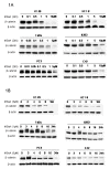
Figure 1
Figure 1
MSeA effect on intracellular expression of β-catenin. Six human cancer cell lines including 2 colorectal (HT-29 and HCT-8), 2 head and neck (FaDu and A253) and 2 prostate (PC3 and C42) were treated with various doses of MSeA (panel 1A) and various times (panel 1B). Western blot analyses show that MSeA inhibits intracellular level of β-catenin in dose and time dependent manners.
Figure 2
Figure 2
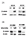
MSeA effect on the nuclear expression of β-catenin. Colorectal cancer cells express a high-level of nuclear β-catenin indicating activation (panel 2A). Treatment with MSeA results in inhibition of the nuclear expression of β-catenin indicating suppression of the active form of β-catenin (panel 2B).
Figure 3
Figure 3
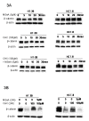
MSeA inhibition of β-catenin is due to degradation. Colorectal cancer cells were treated with a protein synthesis inhibitor, cycloheximide (CHX) and MSeA alone or in combination for various times up to 30 minutes (panel 3A) and for 24 h (panel 3B). No early changes in the β-catenin level were observed (panel 3A). However, 24 h combination treatment with CHX/MSeA resulted in same inhibition after MSeA alone (panel 3B).
Figure 4
Figure 4
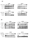
The role of GSK-3β in degradation of β-catenin. Colorectal cancer cells were treated with various doses of MSeA alone (panel 4A) or in combination with GSK-3β inhibitor, Lithium Chloride (LiCl, panel 4B). The level of total GSK-3β, p-GSK-3β and β-catenin were determined using western blots. Treatment with MSeA alone had no effect on total GSK-3β but decreased the level of p-GSK-3β only in HT-29 (panel 4A). Treatments with LiCl increased the level of p-GSK-3β (inactive form of GSK-3β) but had no effect on β-catenin expression (panel 4B). Combination treatment of LiCl/MSeA continued to inhibit expression of β-catenin only in HCT-8 (panel 4A)
Figure 5
Figure 5
Combination treatment effect on the expression of β-catenin. Cancer cells (panel 5A) and nuclear extraction of colorectal cancer cells (panel 5B) were treated with MSeA, SN-38 and docetaxel alone or in combination. Expression of β-catenin was determined using western blots. Combination treatments of MSeA/SN-38 continued to decrease expression of β-catenin when compared with all other groups. Combination treatments of MSeA/docetaxel did not reverse the inhibitory effect of MSeA alone on expression of β-catenin (panel 5A). The active form (nuclear expression) of β-catenin was inhibited by the combination treatment of MSeA/SN-38 when compared with other group (panel 5B).
Figure 6
Figure 6
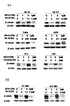
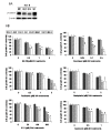
The effect of various drug treatments on cell growth and proliferation in β-catenin knockout cells. Beta-catenin was silenced in colorectal cancer cells (HCT-8, panel 6A). HCT-8 wild type cells (HCT-8WT), scramble controls (HCT-8SC) and 2 β-catenin knockout clones (HCT-8RH7 and HCT-8RF4) were tested. Expression of β-catenin was significantly lower in HCT-8RH1 and HCT-8RF4 when compared with all other groups (panel 6A). Cancer cell growth and proliferation were determined in HCT-8WT, HCT-8SC, HCT-8RH7 and HCT-8RF4 after treatment with various doses of SN-38, docetaxel, paclitaxel, oxaliplatin, 5-FU and topotecan using SRB assay. HCT-8RH7 and HCT-RF4 cells were significantly more sensitive to various drug treatments when compared with HCT-8WT and HCT-8SC (panel 6B). * denotes a p value of less than 0.05 when compared with all other groups.

References

    1. Giles RH, van Es JH, Clevers H. Caught up in a Wnt storm: Wnt signaling in cancer. Biochim Biophys Acta. 2003;1653:1–24. - PubMed
    1. Yang F, Zeng Q, Yu G, Li S, Wang CY. Wnt/beta-catenin signaling inhibits death receptor-mediated apoptosis and promotes invasive growth of HNSCC. Cell Signal. 2006;18:679–687. doi: 10.1016/j.cellsig.2005.06.015. - DOI - PubMed
    1. Goto M, Mitra RS, Liu M, Lee J, Henson BS, Carey T, Bradford C, Prince M, Wang CY, Fearon ER, D'Silva NJ. Rap1 stabilizes beta-catenin and enhances beta-catenin-dependent transcription and invasion in squamous cell carcinoma of the head and neck. Clin Cancer Res. 2010;16:65–76. doi: 10.1158/1078-0432.CCR-09-1122. - DOI - PMC - PubMed
    1. Tsai YP, Yang MH, Huang CH, Chang SY, Chen PM, Liu CJ, Teng SC, Wu KJ. Interaction between HSP60 and beta-catenin promotes metastasis. Carcinogenesis. 2009;30:1049–1057. doi: 10.1093/carcin/bgp087. - DOI - PubMed
    1. Gerstein AV, Almeida TA, Zhao G, Chess E, Shih Ie M, Buhler K, Pienta K, Rubin MA, Vessella R, Papadopoulos N. APC/CTNNB1 (beta-catenin) pathway alterations in human prostate cancers. Genes Chromosomes Cancer. 2002;34:9–16. doi: 10.1002/gcc.10037. - DOI - PubMed

Publication types

LinkOut - more resources