

Incidental cardiac findings on computed tomography imaging of the thorax
- PMID: 21126380
- PMCID: PMC3003672
- DOI: 10.1186/1756-0500-3-326
Incidental cardiac findings on computed tomography imaging of the thorax
Abstract
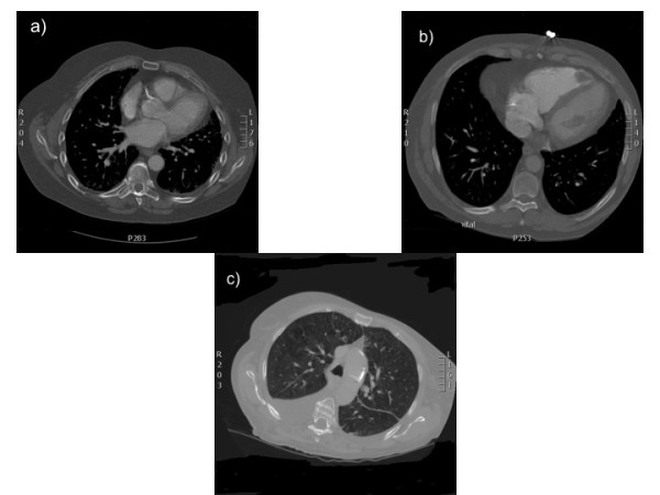
Background: Investigation of pulmonary pathology with computed tomography also allows visualisation of the heart and major vessels. We sought to explore whether clinically relevant cardiac pathology could be identified on computed tomography pulmonary angiograms (CTPA) requested for the exclusion of pulmonary embolism (PE). 100 consecutive CT contrast-enhanced pulmonary angiograms carried out for exclusion of PE at a single centre were assessed retrospectively by two cardiologists.
Findings: Evidence of PE was reported in 5% of scans. Incidental cardiac findings included: aortic wall calcification (54%), coronary calcification (46%), cardiomegaly (41%), atrial dilatation (18%), mitral annulus calcification (15%), right ventricular dilatation (11%), aortic dilatation (8%) and right ventricular thrombus (1%). Apart from 3 (3%) reports describing cardiomegaly, no other cardiac findings were described in radiologists' reports. Other reported pulmonary abnormalities included: lung nodules (14%), lobar collapse/consolidation (8%), pleural effusion (2%), lobar collapse/consolidation (8%), emphysema (6%) and pleural calcification (4%).
Conclusions: CTPAs requested for the exclusion of PE have a high yield of cardiac abnormalities. Although these abnormalities may not have implications for acute clinical management, they may, nevertheless, be important in long-term care.
Figures
References
-
- Garg K, Welsh CH, Feyerabend AJ, Subber SW, Russ PD, Johnston RJ. et al.Pulmonary embolism: diagnosis with spiral CT and ventilation-perfusion scanning - correlation with pulmonary angiographic results or clinical outcome. Radiology. 1998;208:201–208. - PubMed
-
- Perrier A, Howarth N, Didier D, Loubeyre P, Unger PF, de Moerloose P. et al.Performance of helical computed tomography in unselected outpatients with suspected pulmonary embolism. Ann Intern Med. 2001;135:88–97. - PubMed
-
- Mayo JR, Remy-Jardin M, Müller NL, Remy J, Worsley DF, Hossein-Foucher C. et al.Pulmonary embolism: prospective comparison of spiral CT with ventilation-perfusion scintigraphy. Radiology. 1997;205:447–452. - PubMed
-
- Aglan I, Jodocy D, Hiehs S, Soegner P, Frank R, Haberfellner B, Clinical relevance and scope of accidental extracoronary findings in coronary computed tomography angiography: A cardiac versus thoracic FOV study. Eur J Radiol. 2009. - PubMed
LinkOut - more resources
Full Text Sources
