Review
. 2011 Jun 3;711(1-2):87-99.
doi: 10.1016/j.mrfmmm.2010.11.002. Epub 2010 Nov 30.

Mechanism of cluster DNA damage repair in response to high-atomic number and energy particles radiation

Affiliations
Review

Mechanism of cluster DNA damage repair in response to high-atomic number and energy particles radiation

Aroumougame Asaithamby et al. Mutat Res. .

Abstract

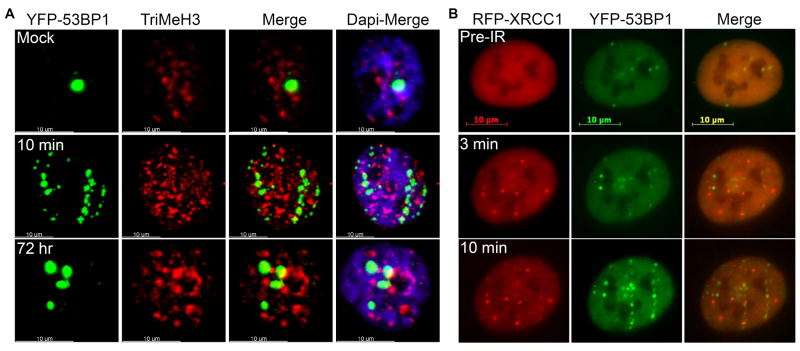
Low-linear energy transfer (LET) radiation (i.e., γ- and X-rays) induces DNA double-strand breaks (DSBs) that are rapidly repaired (rejoined). In contrast, DNA damage induced by the dense ionizing track of high-atomic number and energy (HZE) particles is slowly repaired or is irreparable. These unrepaired and/or misrepaired DNA lesions may contribute to the observed higher relative biological effectiveness for cell killing, chromosomal aberrations, mutagenesis, and carcinogenesis in HZE particle irradiated cells compared to those treated with low-LET radiation. The types of DNA lesions induced by HZE particles have been characterized in vitro and usually consist of two or more closely spaced strand breaks, abasic sites, or oxidized bases on opposing strands. It is unclear why these lesions are difficult to repair. In this review, we highlight the potential of a new technology allowing direct visualization of different types of DNA lesions in human cells and document the emerging significance of live-cell imaging for elucidation of the spatio-temporal characterization of complex DNA damage. We focus on the recent insights into the molecular pathways that participate in the repair of HZE particle-induced DSBs. We also discuss recent advances in our understanding of how different end-processing nucleases aid in repair of DSBs with complicated ends generated by HZE particles. Understanding the mechanism underlying the repair of DNA damage induced by HZE particles will have important implications for estimating the risks to human health associated with HZE particle exposure.


Figures

Figure 1
Figure 1. Clustered DNA lesions induced by iron particles can be visualized by indirect immunostaining with DNA repair and response proteins
(A) Phosphorylated H2AX and DNA PKcs foci form tracks along the paths traversed by iron particles. Normal human skin fibroblasts were irradiated horizontally with iron particles (1 Gy at 1 GeV/nucleon) and stained with γH2AX and DNA-PKcs (pT2609) antibodies 10 min after irradiation. (B) 53BP1, XRCC1, and OGG1 foci form tracks along the densely ionizing paths traversed by iron particles. HT1080 cells were irradiated horizontally with iron particles (1 Gy at 1 GeV/nucleon) and stained with 53BP1, OGG1, and XRCC1 antibodies 10 min after irradiation.
Figure 2
Figure 2. DNA double-strand breaks induced by a spectrum of HZE particles can be directly monitored in live cells
(A) 53BP1 forms foci along the densely ionizing paths traversed by iron (Fe), silicon (Si) and oxygen (O) particles. HT1080 cells stably expressing yellow fluorescent protein (YFP) tagged 53BP1 were imaged prior to irradiation (Pre-IR) and then exposed to Fe, Si and O particles (1 Gy at 1 GeV/nucleon) and were immediately imaged using a Zeiss fluorescent microscope. (B) YFP-53BP1 foci detected in live cells represent the sites of DNA DSBs. HT1080 cells stably expressing yellow fluorescent protein (YFP) tagged 53BP1 were exposed to Fe, Si, and O particles (1 Gy at 1 GeV/nucleon) and were fixed 10 minutes after irradiation. Subsequently, the cells were subjected to indirect immunofluorescence using γH2AX antibodies. (C) XRCC1 forms foci along the densely ionizing paths traversed by iron (Fe), silicon (Si) and oxygen (O) particles. HT1080 cells stably expressing green fluorescent protein (EGFP) tagged XRCC1 were imaged prior to irradiation (Pre-IR) and then exposed to Fe, Si and O particles (1 Gy at 1 GeV/nucleon) and γ-ray and were immediately imaged using a Zeiss fluorescent microscope.
Figure 3
Figure 3. Spatio-temporal characterization of DNA lesions induced by iron particles
(A) Not all persistent 53BP1 foci are located in regions of heterochromatin. HT1080 cells stably expressing YFP-tagged 53BP1 were irradiated horizontally with iron particles (1 Gy at 1 GeV/nucleon) and immunostained with histone H3 (tri methyl K9) antibody (TriMeH3) 10 min and 72 hours after irradiation, and the images were acquired using confocal microscopy (Zeiss). 100-120 cells for each time point in three independent experiments were examined. (B) Iron particle-induced SSBs are detected earlier than the DSBs in single cells by the live-cell imaging approach. HT1080 cells stably expressing dual fluorescent proteins (i.e., YFP-tagged 53BP1 and RFP-tagged XRCC1) were imaged prior to irradiation (Pre-IR), exposed to iron particles (1 Gy at 1 GeV/nucleon), and immediately imaged using Zeiss fluorescent microscopy.
Figure 4
Figure 4. WRN and Artemis play roles in processing the complex DNA damage induced by iron particles
(A) WRN is recruited to the sites of DNA damage induced by iron particles. WS cells stably expressing EGFP-WRN were exposed to iron particles (1Gy at 1GeV/nucleon) and immunostained with γH2AX antibody 30 minutes after irradiation. (B) WRN is important for the processing of DNA damage induced by iron particles. WS cells and WS cells expressing wild-type WRN were irradiated with iron particles (1GeV/nucleon) and were subjected to a colony formation assay. (C) Artemis is critical for the processing of DNA damage induced by iron particles. Artemis-deficient and wild-type cells were exposed to different doses of iron particles (1GeV/nucleon) and were subjected to a colony formation assay. The error bars represent STDEV calculated from at least two independent experiments.

References

    1. Letaw JR, Silberberg R, Tsao CH. Radiation hazards on space missions. Nature. 1987;330:709–710. - PubMed
    1. George K, Durante M, Wu H, Willingham V, Badhwar G, Cucinotta FA. Chromosome aberrations in the blood lymphocytes of astronauts after space flight. Radiat Res. 2001;156:731–738. - PubMed
    1. Goodhead DT. New radiobiological, radiation risk and radiation protection paradigms. Mutat Res. 2010;687:13–16. - PubMed
    1. Durante M, Cucinotta FA. Heavy ion carcinogenesis and human space exploration. Nat Rev Cancer. 2008;8:465–472. - PubMed
    1. Curtis SB, Letaw JR. Galactic cosmic rays and cell-hit frequencies outside the magnetosphere. Adv Space Res. 1989;9:293–298. - PubMed

Publication types

LinkOut - more resources