. 2011 Feb 11;286(6):4209-15.
doi: 10.1074/jbc.M110.194035. Epub 2010 Dec 2.

Repeat expansion affects both transcription initiation and elongation in friedreich ataxia cells

Affiliations

Repeat expansion affects both transcription initiation and elongation in friedreich ataxia cells

Daman Kumari et al. J Biol Chem. .

Abstract

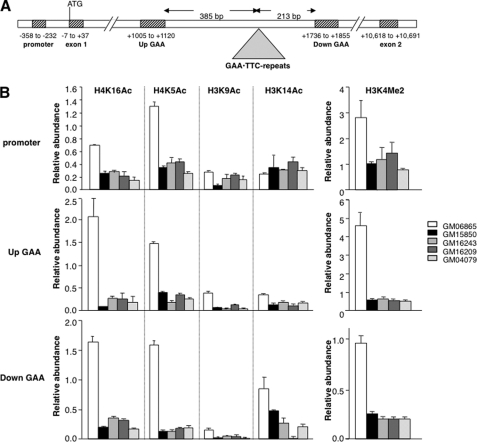
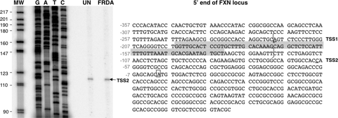
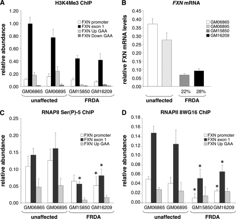
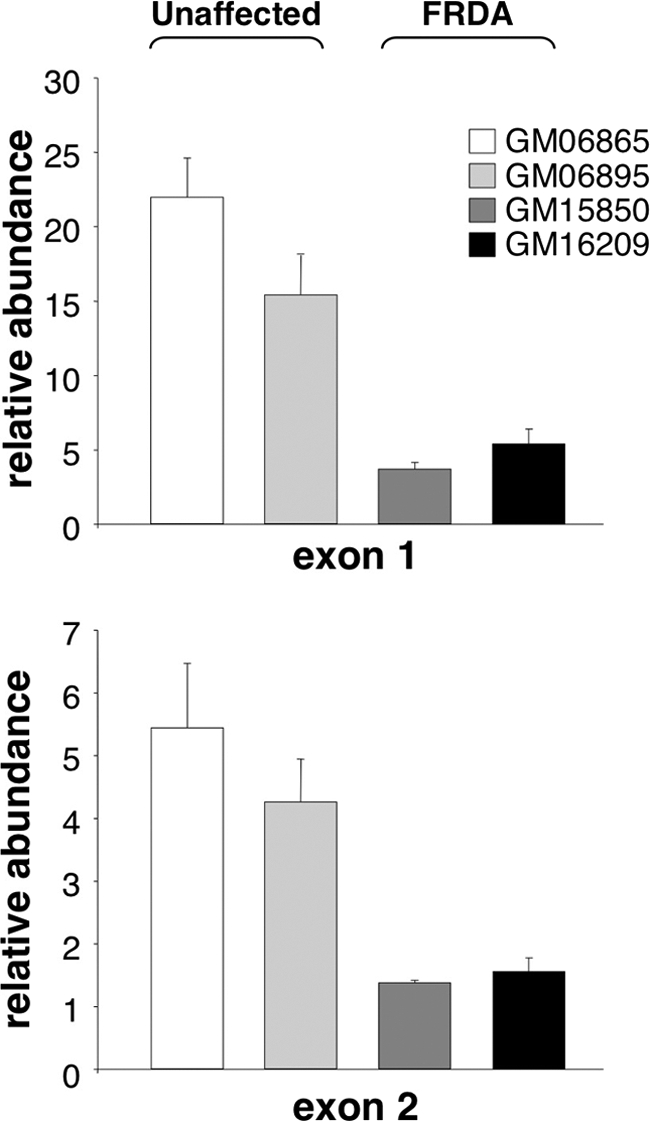
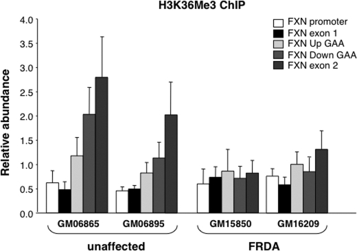
Expansion of a GAA · TTC repeat in the first intron of the frataxin (FXN) gene causes an mRNA deficit that results in Friedreich ataxia (FRDA). The region flanking the repeat on FRDA alleles is associated with more extensive DNA methylation than is seen on normal alleles and histone modifications typical of repressed genes. However, whether these changes are responsible for the mRNA deficit is controversial. Using chromatin immunoprecipitation and cell lines from affected and unaffected individuals, we show that certain marks of active chromatin are also reduced in the promoter region of the FXN gene in patient cells. Thus, the promoter chromatin may be less permissive for transcription initiation than it is on normal alleles. Furthermore, we show that the initiating form of RNA polymerase II and histone H3 trimethylated on lysine 4, a chromatin mark tightly linked to transcription initiation, are both present at lower levels on FRDA alleles. In addition, a mark of transcription elongation, trimethylated H3K36, shows a reduced rate of accumulation downstream of the repeat. Our data thus suggest that repeat expansion reduces both transcription initiation and elongation in FRDA cells. Our findings may have implications for understanding the mechanism responsible for FRDA as well as for therapeutic approaches to reverse the transcription deficit.


Figures

FIGURE 1.
FIGURE 1.
Association of active histone marks with the promoter, Up GAA, and Down GAA region of the frataxin gene in unaffected (GM06865) and FRDA cells (GM15850, GM16243, GM16209, and GM04079). A, schematic representation of the FXN gene showing the positions of the regions analyzed by real-time PCR in ChIP assays. B, chromatin immunoprecipitated with indicated antibodies was normalized to 5% of input and expressed relative to GAPDH. Error bars indicate S.D.
FIGURE 2.
FIGURE 2.
FXN transcription start site in lymphoblastoid cells. Left panel, sequencing gel showing the product of primer extension with end-labeled Exon 1 R2 primer and RNA from unaffected (UN) and FRDA cells using SuperScript III reverse transcriptase according to standard procedures. G, A, T, and C lanes represent the sequencing reactions from the corresponding region of DNA in plasmid FXN81658 (4) using the same primer. MW, pBR322 DNA digested with MspI. Right panel, DNA sequence of the 5′ end of the FXN locus showing the position of previously reported transcription start site, TSS1 (15), and the transcription start site TSS2 identified in this study. The numbers alongside the sequence indicate the position of bases with respect to the first base of the open reading frame, which is shown in the open box. The sequence between the two brackets is the sequence −221 to −121 bases upstream of the frataxin open reading frame, which we showed previously to be important for FXN promoter activity in muscle cells (32). The region highlighted in gray corresponds to the remnants of an ancient L2 repeated DNA element.
FIGURE 3.
FIGURE 3.
Transcription initiation in FRDA cells. A, the abundance of H3K4Me3 on different regions of the FXN gene in unaffected and FRDA cells relative to its abundance on GAPDH. The differences in the levels of H3K4Me3 in unaffected and FRDA cells were only significant for the exon 1 region (p < 0.05). B, FXN mRNA level in unaffected and FRDA cells relative to GUS. Real-time PCR for FXN mRNA was done with primers exon 3 F and exon 4 R as described previously (3). C, relative abundance of DNA immunoprecipitated with an antibody specific to RNAPII phosphorylated at Ser-5 in unaffected and FRDA cells. The asterisks indicate those regions of patient alleles that are significantly different from the corresponding regions of both unaffected alleles tested at p = 0.05 or better. D, relative abundance of DNA immunoprecipitated by the 8WG16 antibody to RNAPII in unaffected and FRDA cells. The asterisks indicate those regions of patient alleles that are significantly different from the corresponding regions of both unaffected alleles tested at p = 0.05 or better. Error bars in all panels indicate S.D.
FIGURE 4.
FIGURE 4.
FXN mRNA levels in unaffected and FRDA cells. The amount of exon 1 and exon 2 was determined using strand-specific PCR as described under “Experimental Procedures.” The levels of FXN mRNA containing exon 1 (upper panel) and exon 2 (lower panel) are shown relative to GUS. Error bars indicate S.D.
FIGURE 5.
FIGURE 5.
The abundance of H3K36Me3 on different regions of the FXN gene in unaffected and FRDA cells. The amount of DNA immunoprecipitated with an antibody to H3K36Me3 was determined as described under “Experimental Procedures” and is shown relative to GAPDH. Error bars indicate S.D.

References

    1. Pandolfo M. (2002) Adv. Exp. Med. Biol. 516, 99–118 - PubMed
    1. Montermini L., Richter A., Morgan K., Justice C. M., Julien D., Castellotti B., Mercier J., Poirier J., Capozzoli F., Bouchard J. P., Lemieux B., Mathieu J., Vanasse M., Seni M. H., Graham G., Andermann F., Andermann E., Melançon S. B., Keats B. J., Di Donato S., Pandolfo M. (1997) Ann. Neurol. 41, 675–682 - PubMed
    1. Herman D., Jenssen K., Burnett R., Soragni E., Perlman S. L., Gottesfeld J. M. (2006) Nat. Chem. Biol. 2, 551–558 - PubMed
    1. Greene E., Mahishi L., Entezam A., Kumari D., Usdin K. (2007) Nucleic Acids Res. 35, 3383–3390 - PMC - PubMed
    1. Al-Mahdawi S., Pinto R. M., Ismail O., Varshney D., Lymperi S., Sandi C., Trabzuni D., Pook M. (2008) Hum. Mol. Genet 17, 735–746 - PubMed

Publication types

MeSH terms