. 2011 Feb 18;286(7):5434-45.
doi: 10.1074/jbc.M110.205054. Epub 2010 Dec 2.

Global analysis of Cdc14 phosphatase reveals diverse roles in mitotic processes

Affiliations

Global analysis of Cdc14 phosphatase reveals diverse roles in mitotic processes

Joanna Bloom et al. J Biol Chem. .

Abstract

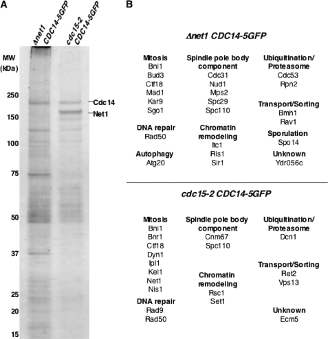
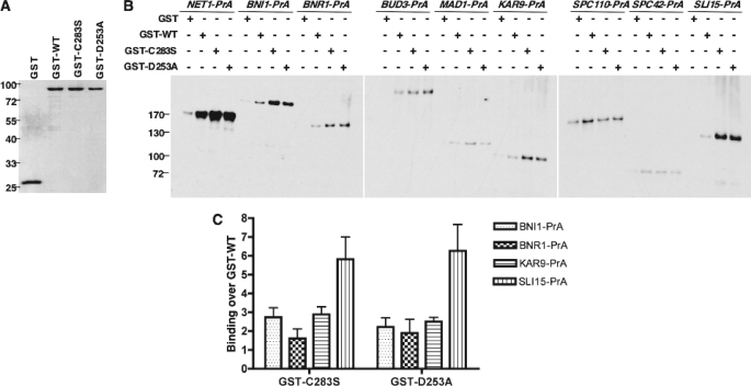
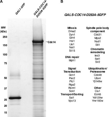
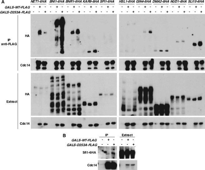
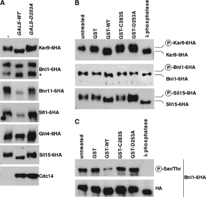
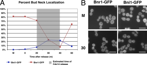
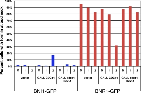
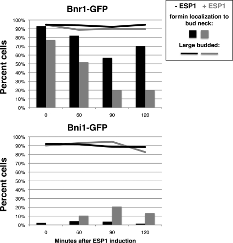
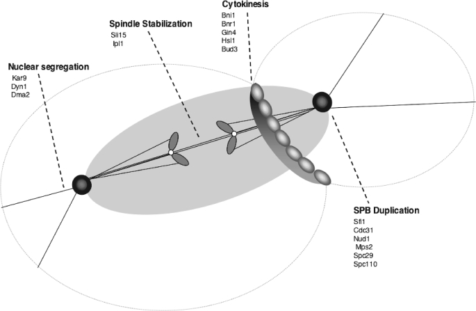
Cdc14 phosphatase regulates multiple events during anaphase and is essential for mitotic exit in budding yeast. Cdc14 is regulated in both a spatial and temporal manner. It is sequestered in the nucleolus for most of the cell cycle by the nucleolar protein Net1 and is released into the nucleus and cytoplasm during anaphase. To identify novel binding partners of Cdc14, we used affinity purification of Cdc14 and mass spectrometric analysis of interacting proteins from strains in which Cdc14 localization or catalytic activity was altered. To alter Cdc14 localization, we used a strain deleted for NET1, which causes full release of Cdc14 from the nucleolus. To alter Cdc14 activity, we generated mutations in the active site of Cdc14 (C283S or D253A), which allow binding of substrates, but not dephosphorylation, by Cdc14. Using this strategy, we identified new interactors of Cdc14, including multiple proteins involved in mitotic events. A subset of these proteins displayed increased affinity for catalytically inactive mutants of Cdc14 compared with the wild-type version, suggesting they are likely substrates of Cdc14. We have also shown that several of the novel Cdc14-interacting proteins, including Kar9 (a protein that orients the mitotic spindle) and Bni1 and Bnr1 (formins that nucleate actin cables and may be important for actomyosin ring contraction) are specifically dephosphorylated by Cdc14 in vitro and in vivo. Our findings suggest the dephosphorylation of the formins may be important for their observed localization change during exit from mitosis and indicate that Cdc14 targets proteins involved in wide-ranging mitotic events.


Figures

FIGURE 1.
FIGURE 1.
Identification of proteins associated with released versus sequestered Cdc14. A, released Cdc14–5GFP from Δnet1 CDC14–5GFP cells or sequestered Cdc14–5GFP from cdc15–2 CDC14–5GFP cells was immunopurified with a polyclonal GFP antibody conjugated to magnetic beads. Eluates were resolved on SDS-PAGE gels and stained with Coomassie Blue. B, Cdc14-associated proteins identified from the gel shown in A were classified into functional groups.
FIGURE 2.
FIGURE 2.
A subset of Cdc14 interactors display enhanced binding to catalytically inactive Cdc14 mutants in vitro. A, Sepharose beads were coated with similar amounts of recombinant GST alone, GST-tagged wild-type Cdc14 (GST-WT) or GST-tagged catalytically inactive Cdc14 mutants (GST-C283S and GST-D253A). Eluates were resolved on SDS-PAGE gels and stained with Coomassie Blue. B, beads shown in A were incubated with extract from the indicated yeast strains. Affinity purified proteins were resolved on SDS-PAGE gels and immunoblotted with a rabbit IgG antibody. C, quantification of binding of Bni1-PrA, Bnr1-PrA, Kar9-PrA, and Sli15-PrA to GST-C283S and GST-D253A over binding to GST-WT. Quantification represents three independent experiments.
FIGURE 3.
FIGURE 3.
Identification of proteins associated with a “substrate-trapping” Cdc14 mutant. A, GFP alone from a control strain (GAL1-GFP) or of “substrate-trapping” Cdc14-D253A-5GFP expressed from a galactose-inducible promoter (GALS-CDC14-D253A-5GFP) was immunopurified with a polyclonal GFP antibody conjugated to magnetic beads. Eluates were resolved on SDS-PAGE gels and stained with Coomassie blue. B, Cdc14-associated proteins identified from the gel shown in A were classified into functional groups.
FIGURE 4.
FIGURE 4.
A subset of Cdc14 interactors display enhanced binding to a substrate-trapping Cdc14 mutant in vivo. A, immunopurification of FLAG-tagged Cdc14 from strains transiently expressing wild-type Cdc14 (GALS-WT-FLAG) or the substrate-trapping Cdc14 mutant (GALS-D253A-FLAG) in combination with the indicated HA6-tagged proteins (upper panels) and whole cell extract from these strains (lower panels). Immunoprecipitates and extracts were resolved on SDS-PAGE gels and immunoblotted with an antibody to the HA tag or to Cdc14. B, immunopurifications of FLAG-tagged Cdc14 from strains transiently expressing wild-type Cdc14 (GALS-WT-FLAG) or the substrate-trapping Cdc14 mutant (GALS-D253A-FLAG) in combination with the indicated Sfi1-HA6 (left panel) and whole cell extract from these strains (right panel). Lysate was extracted using buffer with additional salt and detergent (100 mm NaCl and 0.1% Triton X-100). Immunoprecipitates and extracts were resolved on SDS-PAGE gels and immunoblotted with an antibody to the HA tag or to Cdc14.
FIGURE 5.
FIGURE 5.
Confirmation of new Cdc14 substrates in vivo and in vitro. A, immunoblot analysis of the indicated HA6-tagged proteins in the absence of overexpressed Cdc14 (−) or following transient expression of wild-type Cdc14 (GALS-WT) or the “substrate-trapping” Cdc14 mutant (GALS-D253A). The asterisk indicates a Bni1-HA6 degradation product. B, in vitro phosphatase assay after immunopurification of HA6-tagged proteins following transient expression of GAL-CLB2. Purified proteins were incubated with buffer (−), recombinant GST alone (GST), GST-tagged wild-type Cdc14 (GST-WT), GST-tagged catalytically inactive Cdc14 mutants (GST-C283S and GST-D253A), or 800 U λ phosphatase. Immunoprecipitates were analyzed by immunoblotting with an antibody to HA. C, in vitro phosphatase assay after immunopurification of HA6-tagged Bni1 following transient expression of GAL-CLB2 as in B. Immunoprecipitates were analyzed by immunoblotting with an antibody to phosphoserine/phosphothreonine residues or an antibody to HA.
FIGURE 6.
FIGURE 6.
The timing of the switch in formin localization is coincident with Cdc14 release. A, after a metaphase arrest (M), cells were released synchronously, and the localization of the formins (Bni1 or Bnr1) at the bud neck was scored. The timing of Bnr1 leaving the bud neck is coincident with Bni1 arriving. This switch is also coincident with the timing of Cdc14 release from the nucleolus during the same protocol, gray box (Footnote 7). B, representative images from the experiment in A. Scale bar is 10 μm.
FIGURE 7.
FIGURE 7.
Localization of formins during Cdc14 overexpression at a metaphase arrest. Strains were arrested in metaphase and then galactose was added to induce the expression of CDC14 from the plasmids (vector control, wild type CDC14, or the catalytically inactive cdc14D253A mutant). Presence of the formin at the bud neck is shown during arrest (M) and at 1 and 2 h after addition of galactose (1, 2). Whereas results show CDC14 overexpression caused a relocalization of both formins, further examination showed that the overexpression was also causing exit from mitosis (assayed by actomyosin ring breakdown and DNA content; data not shown).
FIGURE 8.
FIGURE 8.
Cells sustained in anaphase with Cdc14 release show a coincident relocalization of the formins. MET-CDC20 cdh1 GAL-ESP1 strain was arrested in metaphase and either induced with ESP1 (+, in gray) to allow Cdc14 release with delayed cell cycle progression or not (−, black). The localization of the formin to the bud neck at timepoints after the induction is shown for Bnr1-GFP (top panel) and Bni-GFP (bottom panel) by bar graph. To assay cell cycle progression, the percent of large budded cells is shown by the line graphs.
FIGURE 9.
FIGURE 9.
Processes regulated by Cdc14. Potential substrates of Cdc14, identified in this study, are indicated for each mitotic process. Cdc14 has roles in segregation of nuclei, by generating pulling forces on the mother cell-bound SPB, stabilization of the mitotic spindle, by directing proteins to the spindle midzone, and cytokinesis, by influencing cytokinesis and septin ring organization. Cdc14 also has a potential role in licensing of SPB duplication.

References

    1. Schwab M., Lutum A. S., Seufert W. (1997) Cell 90, 683–693 - PubMed
    1. Visintin R., Craig K., Hwang E. S., Prinz S., Tyers M., Amon A. (1998) Mol. Cell 2, 709–718 - PubMed
    1. Jaspersen S. L., Charles J. F., Tinker-Kulberg R. L., Morgan D. O. (1998) Mol. Biol. Cell 9, 2803–2817 - PMC - PubMed
    1. Shou W., Seol J. H., Shevchenko A., Baskerville C., Moazed D., Chen Z. W., Jang J., Shevchenko A., Charbonneau H., Deshaies R. J. (1999) Cell 97, 233–244 - PubMed
    1. Visintin R., Hwang E. S., Amon A. (1999) Nature 398, 818–823 - PubMed

Publication types

MeSH terms