. 2011 Feb 11;286(6):4216-25.
doi: 10.1074/jbc.M110.200295. Epub 2010 Dec 2.

Class II phosphoinositide 3-kinase regulates exocytosis of insulin granules in pancreatic beta cells

Affiliations

Class II phosphoinositide 3-kinase regulates exocytosis of insulin granules in pancreatic beta cells

Veronica Dominguez et al. J Biol Chem. .

Abstract

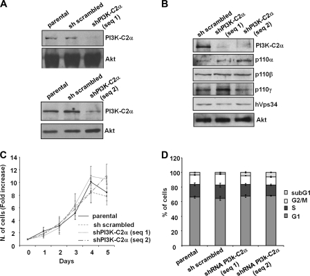
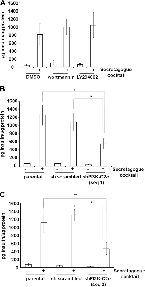
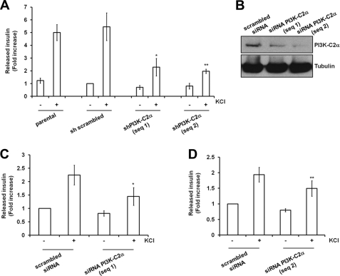
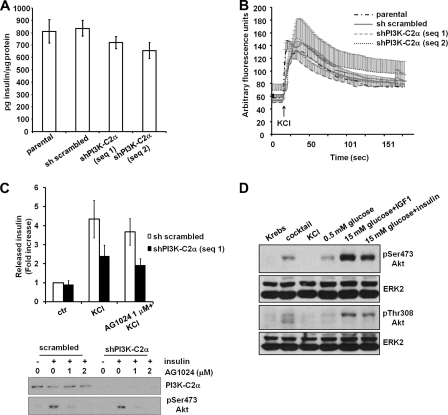
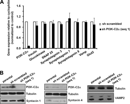
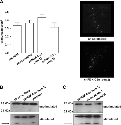
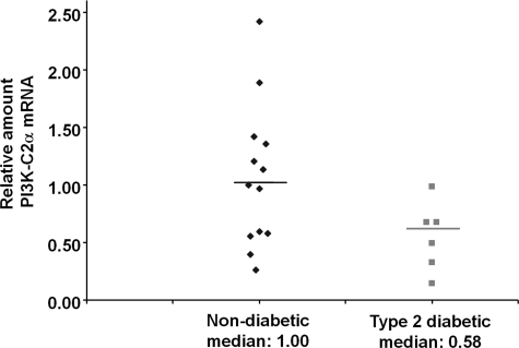
Phosphoinositide 3-kinases (PI3Ks) are critical regulators of pancreatic β cell mass and survival, whereas their involvement in insulin secretion is more controversial. Furthermore, of the different PI3Ks, the class II isoforms were detected in β cells, although their role is still not well understood. Here we show that down-regulation of the class II PI3K isoform PI3K-C2α specifically impairs insulin granule exocytosis in rat insulinoma cells without affecting insulin content, the number of insulin granules at the plasma membrane, or the expression levels of key proteins involved in insulin secretion. Proteolysis of synaptosomal-associated protein of 25 kDa, a process involved in insulin granule exocytosis, is impaired in cells lacking PI3K-C2α. Finally, our data suggest that the mRNA for PI3K-C2α may be down-regulated in islets of Langerhans from type 2 diabetic compared with non-diabetic individuals. Our results reveal a critical role for PI3K-C2α in β cells and suggest that down-regulation of PI3K-C2α may be a feature of type 2 diabetes.


Figures

FIGURE 1.
FIGURE 1.
Generation and characterization of stable INS1 cell lines. A and B, expression levels of PI3K-C2α and other PI3K isoforms in parental INS1 and stable INS1 cells expressing a scrambled shRNA or shRNAs targeting PI3K-C2α are shown. Equal loading was assessed using an anti-Akt antibody (representative blot). C and D, proliferation (C) and cell cycle profile (D) of the indicated cells is shown. Data are the means ± S.E. from three (C) and five (D) independent experiments.
FIGURE 2.
FIGURE 2.
Effect of PI3K-C2α down-regulation on insulin secretion induced by a secretagogue mixture. A, INS1 cells were pretreated for 15 min with 100 nm wortmannin, 10 μm LY294002, or vehicle alone (DMSO), and insulin secretion was determined upon stimulation with a secretagogue mixture for 3 h in the presence of the indicated inhibitors. B and C, insulin secretion was determined upon stimulation of the indicated cell lines with a secretagogue mixture for 3 h. Data are the means ± S.E. from 4 (A), 10 (B), and 3 (C) independent experiments performed in duplicate and are expressed as pg of insulin normalized by total protein content. Student's t test: *, p < 0.01; **, p < 0.05. shPI3K-C2α sequence 1 unstimulated, p < 0.01 versus parental or scrambled unstimulated. shPI3K-C2α sequence 2 unstimulated, not significant versus parental unstimulated or scrambled unstimulated.
FIGURE 3.
FIGURE 3.
Effect of PI3K-C2α down-regulation on insulin secretion induced by treatment with KCl. A, insulin secretion was determined upon stimulation of the indicated stable cell lines with KCl for 30 min. Data are the means ± S.E. from 7 independent experiments performed in duplicate except for shPI3K-C2α sequence 2 (n = 6). Total secreted insulin for each sample (pg insulin) was normalized by the total protein content (μg of protein) and then expressed as -fold increase over the unstimulated sh scrambled cells (pg insulin/μg of protein: 62.467 ± 12.154). Student's t test: *, p < 0.01; **, p < 0.05 versus scrambled or parental. B, shown is Western blotting analysis of PI3K-C2α expression in cells transiently transfected with a scrambled siRNA or siRNAs specifically targeting PI3K-C2α. C and D, insulin secretion in cell lines transiently transfected with the indicated siRNA and stimulated with KCl for 30 min normalized to total protein contents is shown. Data are expressed as -fold increase over insulin released by unstimulated cells expressing the scrambled siRNA (pg insulin/μg protein, 68.145 ± 26.263 for C; pg insulin/μg of protein, 65.385 ± 25.561 for D) and are the means ± S.E. from five independent experiments performed in duplicate. Student's t test: *, p < 0.01; **, p < 0.05.
FIGURE 4.
FIGURE 4.
Investigation of the mechanisms of PI3K-C2α-dependent insulin secretion. A, total insulin content in the indicated cells is shown. Data are the means ± S.E. from nine independent measurements. B, intracellular calcium increase upon treatment of the indicated cells with KCl (in KRB containing 1 mm Ca2+), monitored as specified under “Experimental Procedures,” is shown. Data are the means ± S.E. from 6 independent experiments except for shPI3K-C2α sequence 2 (n = 3). C, cells untreated or pretreated with 1 μm insulin receptor inhibitor AG1024 were left unstimulated or stimulated with KCl for 30 min in the presence or absence of AG1024. Data are the means ± S.E. from five independent experiments performed in duplicate. Total secreted insulin for each sample (pg insulin) was normalized by the total cell number and then expressed as -fold increase over the unstimulated sh scrambled cells (pg insulin/cell: 0.013 ± 0.005). Student's t test KCl-stimulated shPI3K-C2α sequence 1, p < 0.05 versus KCl-stimulated scrambled. AG1024-treated, KCl-stimulated scrambled, not significant versus KCl-stimulated scrambled. AG1024-treated, KCl-stimulated shPI3K-C2α sequence 1, not significant versus KCl-stimulated shPI3K-C2α sequence 1. Blot, cells pretreated for 30 min with the indicated concentrations of AG1024 were stimulated with 100 nm insulin for further 30 min in the presence or absence of AG1024, lysed, and analyzed by Western blot. Akt activation was then determined by analyzing the phosphorylation of its residue Ser-473. D, Western blotting analysis of Akt activation in sh scrambled INS1 was performed as described under “Experimental Procedures.” Equal loading was assessed using an anti-ERK2 antibody.
FIGURE 5.
FIGURE 5.
Down-regulation of PI3K-C2α does not affect expression levels of key proteins involved in insulin secretion. A, results from qPCR analysis of mRNAs extracted from the indicated cells in growing conditions are shown. Data are the means ± S.E. from six preparations. B, shown is a representative Western blot analysis of the levels of syntaxin 1A, syntaxin 4, and VAMP2 in the indicated cells. Corresponding levels of PI3K-C2α are also shown together with tubulin (as loading control).
FIGURE 6.
FIGURE 6.
PI3K-C2α is required for fusion of the insulin granules to the plasma membrane. A, shown is the number of granules on the cell surface as detected by live cell TIRF microscopy. Data are the means ± S.E. from 3–4 independent experiments (n = 23–30 cells per condition). Representative images are shown (average of 4 × 50-ms exposures). Scale bar = 2 μm. B and C, Western blotting analysis of SNAP25 in the indicated unstimulated cells or cells stimulated with the secretagogue mixture for 3h. Arrows indicate the product of proteolysis induced by cellular stimulation.
FIGURE 7.
FIGURE 7.
PI3K-C2α mRNA is altered in islets in type 2 diabetes. Real-time qPCR analysis of PI3K-C2α mRNA levels in human pancreatic islets from non-diabetic (13 samples) compared with type 2 diabetic individuals (6 samples). mRNAs levels were normalized to the reference gene actin. Data are expressed as -fold change relative to the median of the non-diabetic samples. Mann-Whitney's U test one-tailed p < 0.05.

References

    1. Cantley L. C. (2002) Science 296, 1655–1657 - PubMed
    1. Leibiger I. B., Leibiger B., Moede T., Berggren P. O. (1998) Mol. Cell 1, 933–938 - PubMed
    1. Kulkarni R. N., Brüning J. C., Winnay J. N., Postic C., Magnuson M. A., Kahn C. R. (1999) Cell 96, 329–339 - PubMed
    1. Da Silva Xavier G., Qian Q., Cullen P. J., Rutter G. A. (2004) Biochem. J. 377, 149–158 - PMC - PubMed
    1. Zawalich W. S., Zawalich K. C. (2000) J. Endocrinol. 166, 111–120 - PubMed

Publication types

MeSH terms