. 2011 Feb 11;286(6):4292-301.
doi: 10.1074/jbc.M110.189555. Epub 2010 Dec 2.

Molecular basis of BACH1/FANCJ recognition by TopBP1 in DNA replication checkpoint control

Affiliations

Molecular basis of BACH1/FANCJ recognition by TopBP1 in DNA replication checkpoint control

Charles Chung Yun Leung et al. J Biol Chem. .

Abstract

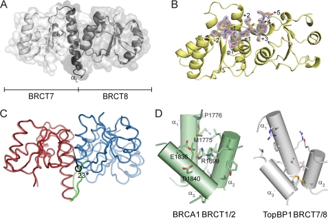
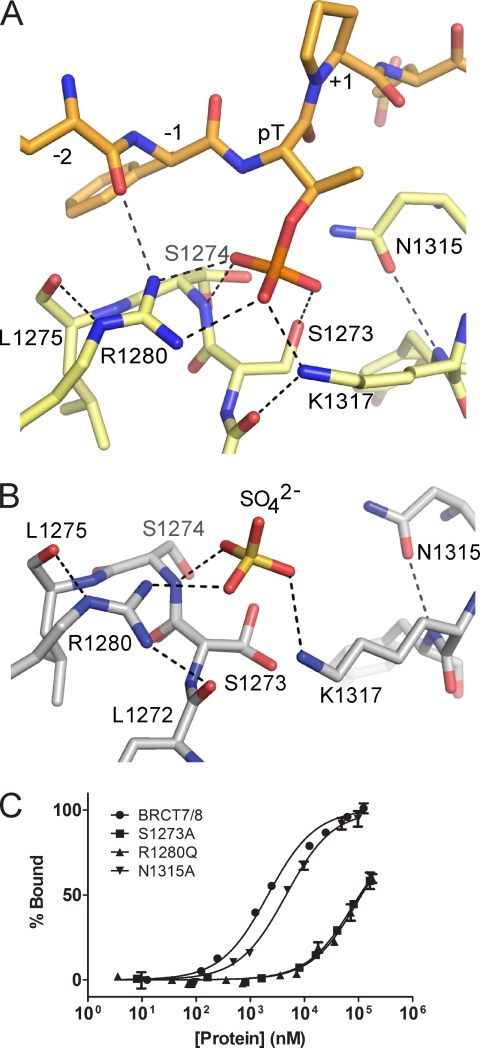
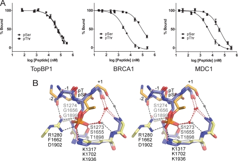
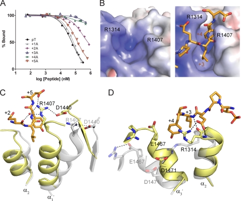
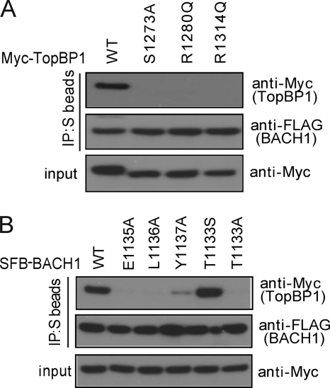
The diverse roles of TopBP1 in DNA replication and checkpoint signaling are associated with the scaffolding ability of TopBP1 to initiate various protein-protein interactions. The recognition of the BACH1/FANCJ helicase by TopBP1 is critical for the activation of the DNA replication checkpoint at stalled replication forks and is facilitated by the C-terminal tandem BRCT7/8 domains of TopBP1 and a phosphorylated Thr(1133) binding motif in BACH1. Here we provide the structural basis for this interaction through analysis of the x-ray crystal structures of TopBP1 BRCT7/8 both free and in complex with a BACH1 phospho-peptide. In contrast to canonical BRCT-phospho-peptide recognition, TopBP1 BRCT7/8 undergoes a dramatic conformational change upon BACH1 binding such that the two BRCT repeats pivot about the central BRCT-BRCT interface to provide an extensive and deep peptide-binding cleft. Additionally, we provide the first structural mechanism for Thr(P) recognition among BRCT domains. Together with systematic mutagenesis studies, we highlight the role of key contacts in governing the unique specificity of the TopBP1-BACH1 interaction.


Figures

FIGURE 1.
FIGURE 1.
Structure of TopBP1 BRCT7/8 and TopBP1 BRCT7/8-BACH1 peptide complex. A, cartoon representation of the apo structure of TopBP1 BRCT7/8. The linker region is colored black, and the αL helix is labeled. B, the BACH1 phospho-peptide (orange) binds in a region spanning the TopBP1 BRCT7/8 domains (yellow). The (2FoFc) electron density map at 2σ for the phospho-peptide is shown. The residue positions in the phospho-peptide are labeled. C, representation of the structural rearrangement of TopBP1 BRCT7/8 around the central rotation axis. The initial apo state is represented at 50% transparency. The fixed domain (red), moving domain (blue), and interdomain bending residues (green) are colored. D, comparison of the hydrophobic packing interface of BRCA1 BRCT1/2 and TopBP1 BRCT7/8. The helices are represented as cylinders and labeled. Residues involved in interface packing are shown as sticks. Residues involved in BRCA1 and not in TopBP1 BRCT packing are labeled.
FIGURE 2.
FIGURE 2.
Phosphate-binding pocket of TopBP1 BRCT7/8. A, stick representation of the TopBP1 BRCT7/8 (yellow) in complex with the BACH1 phospho-peptide (orange). Hydrogen bonding and electrostatic interactions are indicated by dotted lines. B, phosphate-binding pocket of apo TopBP1 BRCT7/8. C, FP binding results for TopBP1 BRCT7/8 phosphate-binding pocket mutants. GST fusion BRCT7/8 variants were purified and used in the assay. Triplicate data points are represented as the means ± S.E.
FIGURE 3.
FIGURE 3.
TopBP1 BRCT7/8 has specificity for Thr(P)- and Ser(P)-binding motifs. A, FP competition assays in which cognate FITC-labeled phospho-peptide complexes of TopBP1 BRCT7/8, BRCA1 BRCT1/2, and MDC1 BRCT1/2 are challenged with their respective Thr(P) and Ser(P) peptides. B, stereo view of the superimposed Ser(P) peptide-specific coordination of BRCA1 (blue, Protein Data Bank code 1T15) and MDC1 (gray, Protein Data Bank code 2AZM) with Thr(P) peptide-specific coordination of TopBP1 (orange-yellow). The residues are labeled for TopBP1 (top), BRCA1 (middle), and MDC1 (bottom). Hydrogen bonding and electrostatic interactions in the complex are represented as dotted lines for TopBP1 (black) and BRCA1/MDC1 (red). Conserved waters mediating peptide-BRCT domain interactions are shown as spheres for TopBP1 (gray) and BRCA1/MDC1 (red).
FIGURE 4.
FIGURE 4.
TopBP1-BACH1 interaction at the +3/+4 binding pocket. A, FP competition analysis of the BACH1-binding motif using alanine scanning mutagenesis. BACH1 phospho-peptides mutated to alanine at +1 to +5 positions were used to compete with the FITC-labeled phospho-peptide bound to TopBP1 BRCT7/8. B, electrostatic potential surface of the TopBP1 BRCT7/8 + 3/+4 binding pocket in the apo (left) and peptide-bound (right) structures. TopBP1 Arg1314 and Arg1407 residues are mapped on the surface. C, role of Arg1407 in the TopBP1-BACH1 complex. TopBP1 BRCT7/8 in the apo (gray) and complex (yellow) structures are superimposed. Residues involved in interacting with Arg1407 are labeled. D, role of Arg1314 in +2/+3 binding of the BACH1 peptide. TopBP1 BRCT7/8 in the apo (gray) and complex (yellow) structures are superimposed.
FIGURE 5.
FIGURE 5.
In vivo binding specificities of the TopBP1-BACH1 interaction. A, effects of TopBP1 BRCT7/8 mutations in binding BACH1. Constructs encoding Myc-tagged wild type and mutants of TopBP1 were co-transfected with plasmids encoding SFB-BACH1. Immunoprecipitation (IP) reactions were performed using S protein beads and then subjected to Western blot analyses using antibodies as indicated. B, effects of BACH1 mutations in binding TopBP1. Lysates for immunoprecipitation were prepared from cells expressing Myc-tagged TopBP1 along with SFB-tagged wild type or mutants of BACH1.

References

    1. Nyberg K. A., Michelson R. J., Putnam C. W., Weinert T. A. (2002) Annu. Rev. Genet. 36, 617–656 - PubMed
    1. Osborn A. J., Elledge S. J., Zou L. (2002) Trends Cell Biol. 12, 509–516 - PubMed
    1. Cimprich K. A., Cortez D. (2008) Nat. Rev. Mol. Cell Biol. 9, 616–627 - PMC - PubMed
    1. Burrows A. E., Elledge S. J. (2008) Genes Dev. 22, 1416–1421 - PMC - PubMed
    1. Gong Z., Kim J. E., Leung C. C., Glover J. N., Chen J. (2010) Mol. Cell 37, 438–446 - PMC - PubMed

Publication types

MeSH terms