. 2010 Dec 3;330(6009):1390-3.
doi: 10.1126/science.1190217.

BID, BIM, and PUMA are essential for activation of the BAX- and BAK-dependent cell death program

Affiliations

BID, BIM, and PUMA are essential for activation of the BAX- and BAK-dependent cell death program

Decheng Ren et al. Science. .

Abstract

Although the proteins BAX and BAK are required for initiation of apoptosis at the mitochondria, how BAX and BAK are activated remains unsettled. We provide in vivo evidence demonstrating an essential role of the proteins BID, BIM, and PUMA in activating BAX and BAK. Bid, Bim, and Puma triple-knockout mice showed the same developmental defects that are associated with deficiency of Bax and Bak, including persistent interdigital webs and imperforate vaginas. Genetic deletion of Bid, Bim, and Puma prevented the homo-oligomerization of BAX and BAK, and thereby cytochrome c-mediated activation of caspases in response to diverse death signals in neurons and T lymphocytes, despite the presence of other BH3-only molecules. Thus, many forms of apoptosis require direct activation of BAX and BAK at the mitochondria by a member of the BID, BIM, or PUMA family of proteins.


Figures

Fig. 1
Fig. 1
Triple deficiency of Bid, Bim, and Puma phenocopies double deficiency of Bax and Bak. (A) Bid-/-Bim-/-Puma-/- TKO mice display persistence of interdigital webs. Ventral views of paws from WT, Bid-/-Bim-/-, and Bid-/-Bim-/-Puma-/- mice. (B) Bid-/-Bim-/-Puma-/- TKO mice fail to develop external vaginal introituses. Photographs of vaginal openings from WT, Bid-/-Bim-/-, and Bid-/-Bim-/-Puma-/- mice. Arrows point to external vaginal region. (C to G) Immunohistochemistry for cleaved caspase-3 from cerebella of postnatal day 5 (P5) mice of the indicated genotypes that were irradiated with 14 Gy γ-irradiation. (C) WT, 18 hours after IR. (D) Baxf/-Bak-/-Nestin-Cre+, 30 hours after IR. (E) Puma-/-, 30 hours after IR. (F) Bim-/-Puma-/-, 30 hours after IR. (G) Bid-/-Bim-/-Puma-/-, 30 hours after IR. Arrows denote the external granular layer of cerebellum. Data shown are representative images of two to three independent experiments. (H) Analyses of a whole sagittal section of cerebellum at the same level from experiments shown in (C to G) summarize the numbers of neurons at the external granular layer that were stained positive for cleaved caspase-3. Data shown are the average of two independent experiments (n = 2).
Fig. 2
Fig. 2
Bid-/-Bim-/-Puma-/- TKO cerebellar granule neurons are completely resistant to potassium-deprivation induced apoptosis. Cerebellar granule neurons from WT, Bim-/-, Puma-/-, Bim-/-Puma-/-, or Bid-/-Bim-/-Puma-/- mice were cultured in high-K+ (K25 + S) for 7 days and then transferred to low-K+ medium (K5 + S) to induce apoptosis. Viability was determined at the indicated times using propidium iodide (PI) staining. Data are the mean percentage of PI-positive neurons ± SD from three independent experiments. **, P < 0.01; ***, P < 0.001.
Fig. 3
Fig. 3
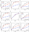
Bid-/-Bim-/-Puma-/- TKO T cells are resistant to diverse apoptotic stimuli. (A to E) CD4+ T cells purified from the spleens of WT (n = 3), Bid-/-Bim-/-(n = 3), or Bid-/-Bim-/-Puma-/- (n = 3) mice at 8 to 10 weeks of age were cultured under the following conditions: in the absence of cytokine (A), after exposure to 2.5 Gy γ-irradiation (B), in the presence of etoposide (C), in the presence of dexamethasone (D), or in the presence of agonistic antibody to Fas (E). Cell death was quantified by annexin-V staining at the indicated times. (F to I) Thymocytes from WT (n = 3), Bid-/-Bim-/-(n = 3), or Bid-/-Bim-/-Puma-/- (n = 3) mice at 6 to 8 weeks of age were cultured under the following conditions: in the absence of cytokine (F), after exposure to 2.5 Gy γ-irradiation (G), in the presence of etoposide (H), or in the presence of tunicamycin (I). Cell death was quantified by annexin-V staining at the indicated times. Data are the mean percentage of annexin-V-positive cells ± SD from three independent experiments. *, P < 0.05; **, P < 0.01; ***, P < 0.001.
Fig. 4
Fig. 4
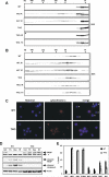
BID, BIM, and PUMA are required to activate BAX- and BAK-dependent mitochondrial apoptosis. (A and B) Triple deficiency of Bid, Bim, and Puma prevents activation of BAX and BAK. Thymocytes from WT or Bid-/-Bim-/-Puma-/- mice were untreated, irradiated with 5 Gy γ-irradiation (IR), or treated with tunicamycin (TC). Protein lysates were harvested at 7 hours after IR or 20 hours after TC treatment and subjected to Superdex 200 (HR10/30) gel-filtration chromatography. Fractions were analyzed by Western blot using antibodies to BAX (A) or BAK (B). (C) Triple deficiency of Bid, Bim, and Puma prevents the translocation of cytochrome c. Fluorescence microscopy of WT or Bid-/-Bim-/-Puma-/- thymocytes 20 hours after exposure to 2.5 Gy γ-irradiation. Red represents cytochrome c immunostaining, and blue is Hoechst staining. White asterisks indicate apoptotic cells that have lost cytochrome c. (D) Triple deficiency of Bid, Bim, and Puma prevents the activation of caspases. Thymocytes from WT or Bid-/-Bim-/-Puma-/- mice were untreated, treated with TC or dexamethasone (Dex), or irradiated with 5 Gy γ-irradiation (IR). After 12 hours, protein lysates were harvested and assessed by Western blot using antibodies specific for PARP, cleaved PARP, cleaved caspase-3, or actin. Asterisk denotes a cross-reactive protein. (E) Primary mouse embryonic fibroblasts isolated from WT or Bid-/-Bim-/-Puma-/- TKO mice were infected with retroviruses expressing the indicated genes. Cell death was quantified by annexin-V staining at 24 hours. Data are the mean percentage of annexin-V-positive cells ± SD from three independent experiments. *, P < 0.05.

Comment in

References

    1. Gross A, McDonnell JM, Korsmeyer SJ. Genes Dev. 1999;13:1899. - PubMed
    1. Wang X. Genes Dev. 2001;15:2922. - PubMed
    1. Newmeyer DD, Ferguson-Miller S. Cell. 2003 Feb 21;112:481. - PubMed
    1. Reed JC. Cell Death Differ. 2006 Aug;13:1378. - PubMed
    1. Youle RJ, Strasser A. Nat Rev Mol Cell Biol. 2008 Jan;9:47. - PubMed

Publication types

MeSH terms