. 2010 Dec 21;107(51):22090-5.
doi: 10.1073/pnas.1009182107. Epub 2010 Dec 2.

Apurinic/apyrimidinic (AP) site recognition by the 5'-dRP/AP lyase in poly(ADP-ribose) polymerase-1 (PARP-1)

Affiliations

Apurinic/apyrimidinic (AP) site recognition by the 5'-dRP/AP lyase in poly(ADP-ribose) polymerase-1 (PARP-1)

S N Khodyreva et al. Proc Natl Acad Sci U S A. .

Abstract

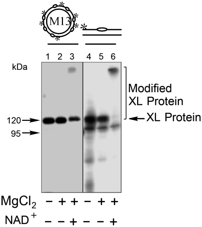
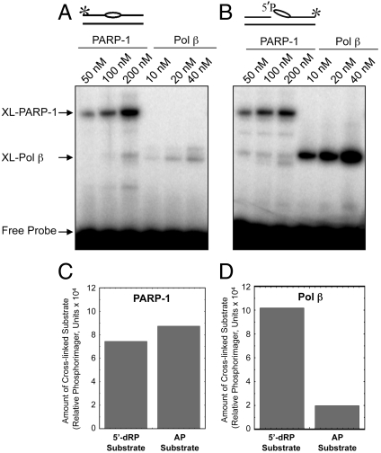
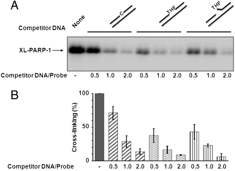
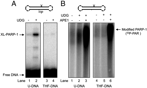
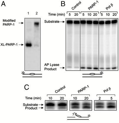
The capacity of human poly(ADP-ribose) polymerase-1 (PARP-1) to interact with intact apurinic/apyrimidinic (AP) sites in DNA has been demonstrated. In cell extracts, sodium borohydride reduction of the PARP-1/AP site DNA complex resulted in covalent cross-linking of PARP-1 to DNA; the identity of cross-linked PARP-1 was confirmed by mass spectrometry. Using purified human PARP-1, the specificity of PARP-1 binding to AP site-containing DNA was confirmed in competition binding experiments. PARP-1 was only weakly activated to conduct poly(ADP-ribose) synthesis upon binding to AP site-containing DNA, but was strongly activated for poly(ADP-ribose) synthesis upon strand incision by AP endonuclease 1 (APE1). By virtue of its binding to AP sites, PARP-1 could be poised for its role in base excision repair, pending DNA strand incision by APE1 or the 5'-dRP/AP lyase activity in PARP-1.


Conflict of interest statement

The authors declare no conflict of interest.

Figures

Fig. 1.
Fig. 1.
Cross-linking of BTNE proteins to AP site-containing DNA probe and effect of poly(ADP-ribose) modification. The reaction conditions and product analysis were as described under Materials and Methods. 32P-labeled AP site-containing circular DNA probe (20 nM) or linear DNA probe (100 nM) was incubated with BTNE (1.25 mg/mL (lanes 2 and 5) for 15 min at 37 °C. In lanes 3 and 6, the reaction mixtures were supplemented with NAD+ and then further incubated for 5 min at 37 °C prior to NaBH4 treatment. NaBH4 was then added, and the mixtures were further incubated at 0–1 °C for 30 min. Then in lane 1, the reaction mixture was supplemented with 10 mM MgCl2. The reaction products in lanes 1–3 (proteins cross-linked with circular AP DNA) were treated with benzonase (12 units) for 15 min at 37 °C prior to SDS-PAGE. Covalently cross-linked DNA-protein complexes (XL Protein) were separated by SDS-PAGE. A representative phosphorimage illustrating cross-linking of BTNE proteins is shown. The migration positions of protein markers (M), designated in kDa, XL Protein, and poly(ADP-ribose) Modified XL Protein are indicated.
Fig. 2.
Fig. 2.
Comparison of cross-linking of purified PARP-1 and Pol β with 5′-dRP lyase substrate DNA and AP site-containing DNA. Cross-linking of purified PARP-1 to AP site-containing linear DNA was performed as described under Materials and Methods. Schematic representations of DNA probes are shown at Top. The * symbol denotes the position of the 32P label in the DNA. The bubble-like symbol denotes the presence of the AP site in the DNA. Representative results of PARP-1 and Pol β cross-linking to intact AP site-containing DNA (A) or preincised AP site-containing DNA (B) as a function of protein concentration. Quantification showing the yield of XL-PARP-1 (C) and XL-Pol β (D). Relative phosphorimager units of cross-linked products formed in the presence of 200 nM PARP-1 (C) and 40 nM Pol β (D) are shown.
Fig. 3.
Fig. 3.
Specificity of the PARP-1 cross-linking with AP site-containing DNA, as revealed by competition with control and synthetic (THF) AP site-containing DNAs. Cross-linking of purified PARP-1 to linear natural AP site-containing DNA and the competition experiments were performed as described under Materials and Methods. (A) A representative phosphorimage showing the results of PARP-1 cross-linked to 32P-labeled AP site-containing DNA in the absence (−) or presence of competitor DNA, as indicated. The competitor-to-probe ratio is indicated. Competitor DNA contained either cytosine (C) or synthetic AP site (THF) at the same position as the natural AP site in the labeled DNA probe. A schematic representation of competitor DNAs is at Top. (B) Quantification summarizing the yield of XL-PARP-1 in three experiments similar to the experiment shown in A. In each independent experiment, the cross-linking in the absence of competitor DNA was taken as 100%. The data are the mean ± SD, n = 3.
Fig. 4.
Fig. 4.
Cross-linking and poly(ADP-ribosyl)ation activities of purified PARP-1. Experiments in A and B were performed with double-hairpin DNA substrates containing a uracil base or THF synthetic AP site, as specified under the gel images. A schematic representation of the hairpin DNA is at Top. The symbol “X” denotes the position of uracil (i.e., natural AP site) or the synthetic AP site in the DNA. (A) Results of purified PARP-1 cross-linking, as a function of UDG treatment, to internal 32P-labeled double-hairpin DNA with a uracil residue or a THF synthetic AP site. Cross-linking of PARP-1 was performed either without (lanes 1 and 3) or with (lanes 2 and 4) UDG treatment, as illustrated and described under Materials and Methods. The positions of XL-PARP-1 and free DNA are indicated. (B) Results of a poly(ADP-ribosyl)ation assay with intact (lanes 1, 2, 4, and 5) and APE1 incised (lanes 3 and 6) AP site DNAs. The presence (+) and absence (−) of UDG is indicated. Poly(ADP-ribosyl)ation was measured after incubations in the presence of 32P-labed NAD. Reaction conditions are described under Materials and Methods.
Fig. 5.
Fig. 5.
Cross-linking, poly(ADP-ribosyl)ation and 5′-dRP/AP lyase activities of purified PARP-1. (A) Cross-linking (lane 1) and poly(ADP-ribosyl)ation (lane 2) were performed as in Fig. 1 using purified PARP-1 and 32P-labeled linear natural AP site-containing DNA. The positions of XL-PARP-1 and poly(ADP-ribose) modified XL-PARP-1 are indicated, and the DNA is illustrated at the bottom. (B) AP lyase activity of purified PARP-1. Reaction mixtures containing 32P-labeled intact AP site-containing DNA were assembled on ice with PARP-1 (200 nM), Pol β (200 nM), or buffer (control). Aliquots were withdrawn at the indicated intervals and analyzed for the strand incision AP lyase activity product as described under Materials and Methods. Lanes 1 and 2 represent AP site stability in the absence of PARP-1. (C) 5′-dRP lyase activity of purified PARP-1. Reaction mixtures with PARP-1 (100 nM) and Pol β (5 nM) were incubated as described. The positions of the substrates and products are indicated, and the DNA is illustrated at the bottom. The * symbol illustrates the position of the 32P label in the DNA, as in Fig. 2. In B, all eight lanes shown were from the same gel; some lanes from this gel are not shown because they are not relevant to the interpretation of this experiment, and the order of the original lanes was adjusted for use in B.

References

    1. Lindahl T. Instability and decay of the primary structure of DNA. Nature. 1993;362:709–715. - PubMed
    1. Wilson DM, 3rd, Thompson LH. Life without DNA repair. Proc Natl Acad Sci USA. 1997;94:12754–12757. - PMC - PubMed
    1. McCullough AK, Dodson ML, Lloyd RS. Initiation of base excision repair: Glycosylase mechanisms and structures. Annu Rev Biochem. 1999;68:255–285. - PubMed
    1. Atamna H, Cheung I, Ames BN. A method for detecting abasic sites in living cells: Age-dependent changes in base excision repair. Proc Natl Acad Sci USA. 2000;97:686–691. - PMC - PubMed
    1. Burrows CJ, Muller JG. Oxidative nucleobase modifications leading to strand scission. Chem Rev. 1998;98:1109–1152. - PubMed

Publication types

MeSH terms