

Strongly bound citrate stabilizes the apatite nanocrystals in bone
- PMID: 21127269
- PMCID: PMC3012505
- DOI: 10.1073/pnas.1009219107
Strongly bound citrate stabilizes the apatite nanocrystals in bone
Abstract
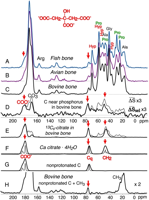
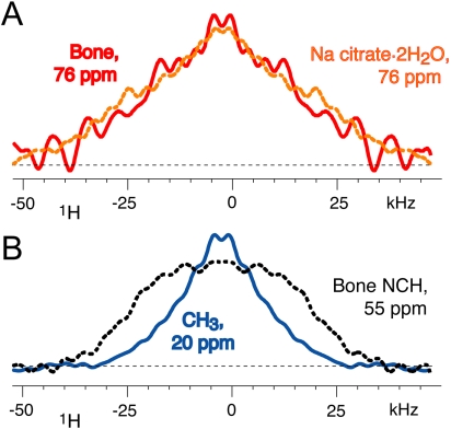
Nanocrystals of apatitic calcium phosphate impart the organic-inorganic nanocomposite in bone with favorable mechanical properties. So far, the factors preventing crystal growth beyond the favorable thickness of ca. 3 nm have not been identified. Here we show that the apatite surfaces are studded with strongly bound citrate molecules, whose signals have been identified unambiguously by multinuclear magnetic resonance (NMR) analysis. NMR reveals that bound citrate accounts for 5.5 wt% of the organic matter in bone and covers apatite at a density of about 1 molecule per (2 nm)(2), with its three carboxylate groups at distances of 0.3 to 0.45 nm from the apatite surface. Bound citrate is highly conserved, being found in fish, avian, and mammalian bone, which indicates its critical role in interfering with crystal thickening and stabilizing the apatite nanocrystals in bone.
Conflict of interest statement
The authors declare no conflict of interest.
Figures
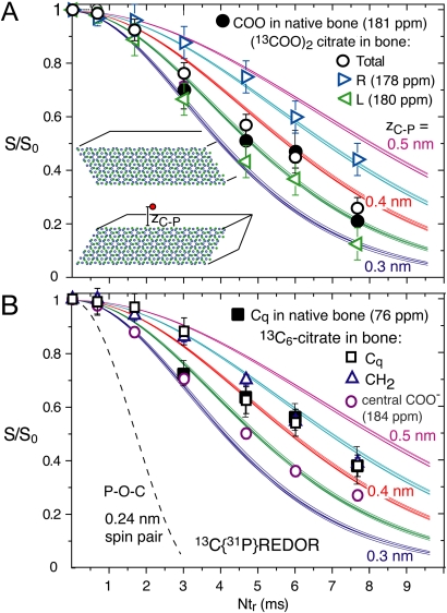
 groups in 13C-labeled citrate absorbed into bovine bone. Triangles pointing right and left: Dephasing of 178- and 180-ppm 13C citrate signals, respectively. (B) Data for the quaternary C-OH of citrate in bone (filled black squares), as well as quaternary C─OH (Cq, open squares), CH2 (open blue triangles) and the central COO- (open purple circles) in 13C-labeled citrates absorbed into bovine bone. Error margins were determined from the signal-to-noise ratios in the REDOR spectra. The dashed curve is for a 31C-31P spin pair (two-bond distance of 0.24 nm) as in phosphocitrate.
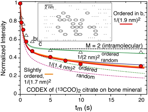
Comment in
-
How to control the size and morphology of apatite nanocrystals in bone.Proc Natl Acad Sci U S A. 2010 Dec 28;107(52):22369-70. doi: 10.1073/pnas.1017493108. Epub 2010 Dec 17. Proc Natl Acad Sci U S A. 2010. PMID: 21169505 Free PMC article. No abstract available.
Similar articles
-
How to control the size and morphology of apatite nanocrystals in bone.Proc Natl Acad Sci U S A. 2010 Dec 28;107(52):22369-70. doi: 10.1073/pnas.1017493108. Epub 2010 Dec 17. Proc Natl Acad Sci U S A. 2010. PMID: 21169505 Free PMC article. No abstract available.
-
Crystallization of bioinspired citrate-functionalized nanoapatite with tailored carbonate content.Acta Biomater. 2012 Sep;8(9):3491-9. doi: 10.1016/j.actbio.2012.04.046. Epub 2012 May 8. Acta Biomater. 2012. PMID: 22579712
-
HRTEM study of individual bone apatite nanocrystals reveals symmetry reduction with respect to P63/m apatite.Mater Sci Eng C Mater Biol Appl. 2019 Nov;104:109966. doi: 10.1016/j.msec.2019.109966. Epub 2019 Jul 9. Mater Sci Eng C Mater Biol Appl. 2019. PMID: 31499942
-
Biomineralization mechanisms: a new paradigm for crystal nucleation in organic matrices.Calcif Tissue Int. 2013 Oct;93(4):307-15. doi: 10.1007/s00223-012-9678-2. Epub 2012 Dec 16. Calcif Tissue Int. 2013. PMID: 23241924 Free PMC article. Review.
-
[Anisotropic crystallization of biominerals].Clin Calcium. 2014 Feb;24(2):193-202. Clin Calcium. 2014. PMID: 24473352 Review. Japanese.
Cited by
-
Calcium orthophosphates: occurrence, properties, biomineralization, pathological calcification and biomimetic applications.Biomatter. 2011 Oct-Dec;1(2):121-64. doi: 10.4161/biom.18790. Biomatter. 2011. PMID: 23507744 Free PMC article. Review.
-
Fast degradable citrate-based bone scaffold promotes spinal fusion.J Mater Chem B. 2015 Jul 21;3(27):5569-5576. doi: 10.1039/C5TB00607D. J Mater Chem B. 2015. PMID: 26213625 Free PMC article.
-
The Role of Hydroxyl Channel in Defining Selected Physicochemical Peculiarities Exhibited by Hydroxyapatite.RSC Adv. 2015;5:36614-36633. doi: 10.1039/C4RA17180B. RSC Adv. 2015. PMID: 26229593 Free PMC article.
-
Development of Injectable Citrate-Based Bioadhesive Bone Implants.J Mater Chem B. 2015 Jan 21;3:387-398. doi: 10.1039/C4TB01498G. J Mater Chem B. 2015. PMID: 25580247 Free PMC article.
-
Mechanistic Insights into the Structural Stability of Collagen-Containing Biomaterials Such as Bones and Cartilage.J Phys Chem B. 2021 May 13;125(18):4757-4766. doi: 10.1021/acs.jpcb.1c01431. Epub 2021 Apr 30. J Phys Chem B. 2021. PMID: 33929847 Free PMC article.
References
-
- Buckwalter JA, Glimcher MJ, Cooper RR, Recker R. Bone biology .1. Structure, blood-supply, cells, matrix, and mineralization. J Bone Joint Surg Am. 1995;77A:1256–1275. - PubMed
-
- Currey JD. Bones. Princeton, New Jersey: Princeton University Press; 2002. p. 456.
-
- Weiner S, Traub W. Bone-structure—from Angstroms to microns. FASEB J. 1992;6:879–885. - PubMed
-
- Weiner S, Wagner HD. The material bone: Structure mechanical function relations. Annu Rev Mater Sci. 1998;28:271–298.
Publication types
MeSH terms
Substances
LinkOut - more resources
Full Text Sources
Other Literature Sources
