. 2010 Dec 10;107(12):1498-502.
doi: 10.1161/CIRCRESAHA.110.233080. Epub 2010 Dec 2.

Endothelial nitric oxide modulates expression and processing of amyloid precursor protein

Affiliations

Endothelial nitric oxide modulates expression and processing of amyloid precursor protein

Susan A Austin et al. Circ Res. .

Abstract

Rationale: the exact etiology of sporadic Alzheimer disease (AD) is unclear, but it is interesting that several cardiovascular risk factors are associated with higher incidence of AD. The link between these risk factors and AD has yet to be identified; however, a common feature is endothelial dysfunction, specifically, decreased bioavailability of nitric oxide (NO).

Objective: to determine the relationship between endothelial derived NO and the expression and processing of amyloid precursor protein (APP).

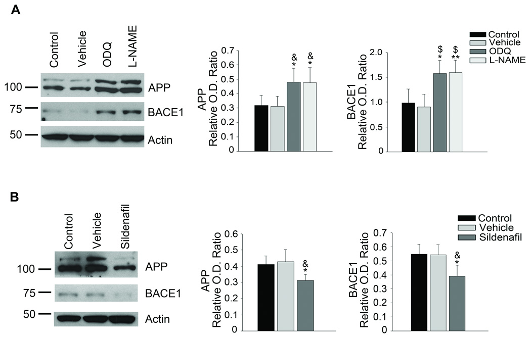
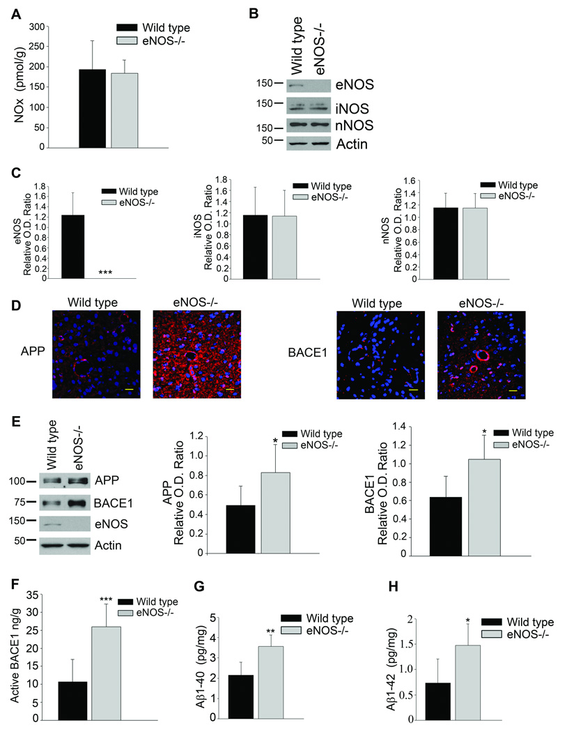
Methods and results: we used human brain microvascular endothelial cells to examine the role of NO in modulating APP expression and processing in vitro. Inhibition of endothelial nitric oxide synthase (eNOS) with the specific NOS inhibitor L-NAME (N(G)-nitro-l-arginine methyl ester) led to increased APP and BACE1 (β-site APP-cleaving enzyme1) protein levels, as well as increased secretion of the amyloidogenic peptide amyloid β (Aβ) (control 10.93 ± 0.70 pg/mL; L-NAME 168.21 ± 27.38 pg/mL; P<0.001). To examine the role of NO in modulation of APP expression and processing in vivo, we used brain and cerebral microvessels from eNOS-deficient (eNOS(-/-)) mice. Brain tissue from eNOS(-/-) mice had statistically higher APP and BACE1 protein levels, as well as increased BACE1 enzyme activity and Aβ (Aβ(1)(-)(42) wild-type control, 0.737 pg/mg; eNOS(-/-), 1.475 pg/mg; P<0.05), compared with wild-type controls (n=6 to 8 animals per background). Brain microvessels from eNOS(-/-) mice also showed statistically higher BACE1 protein levels as compared with wild-type control.

Conclusions: our data suggest that endothelial NO plays an important role in modulating APP expression and processing within the brain and cerebrovasculature. The NO/cGMP pathway may be an important therapeutic target in preventing and treating mild cognitive impairment, as well as AD.


Figures

Figure 1
Figure 1
Inhibitory effect of eNOS-derived NO on APP and BACE1 protein expression and Aβ generation in human BMEC. A, BMEC were cultured in the absence or presence of 0.3 mM L-NAME for 1, 3, and 5 days; medium was changed daily. Representative image from 3–5 independent experiments and densitometric analysis is shown. Data is presented as mean ±SD (*P<0.05 from control, **P<0.01 from control). BMEC were cultured in the absence or presence of 1 mM L-Arginine or D-Arginine and 0.3 mM L-NAME for 4 days; medium was changed daily. B, Representative image from 3–5 independent experiments and densitometric analysis shown. Data is presented as mean ±SD (*P<0.05 from control, &P<0.05 from L-NAME, $P<0.01 from L-NAME). C, Cell supernatant was collected and secreted Aβ1–40 and Aβ1–42 was measured using a commercially available colorimetric ELISA. Data is represented as mean ± SD (***P<0.001 from control, #P<0.001 from L-NAME, $P<0.01 from L-NAME, &P<0.05 from L-NAME). D, BMEC were transfected with 30 nM of eNOS or Control siRNA. Cells were lysed 3 days after transfections and used for Western analyses. Representative image from 3–5 independent experiments and densitometric analysis is shown. Data is presented as mean ±SD (*P<0.05 from untreated, &P<0.05 from Control siRNA). Statistical analysis was performed using one-way ANOVA and Tukey-Kramer post hoc comparison.
Figure 2
Figure 2
NO-mediated suppression of APP and BACE1 protein expression is mediated by the guanylyl cyclase/cGMP pathway. A, BMEC were cultured in the absence or presence of DMSO vehicle or 1 µM ODQ, a soluble guanylyl cyclase inhibitor, for 4 days, medium was changed daily. Representative image from 3–5 independent experiments and densitometric analysis is shown. Data is presented as mean ±SD (*P<0.05 from control, **P<0.01 from control, &P<0.05 from vehicle, $P<0.01 from vehicle). B, BMEC were cultured in the absence or presence of 1 µM sildenafil citrate, for 4 days, medium was changed daily. Representative image from 3–5 independent experiments and densitometric analysis is shown. Data is presented as mean ±SD (*P<0.05 from control, &P<0.05 from vehicle).
Figure 3
Figure 3
Levels of NO were decreased while BACE1 protein levels were increased in the micovessels of eNOS−/− mice as compared to age-matched wild type control animals. A, Brain microvascular tissue from eNOS−/− and wild type animals was collected and analyzed for levels of NOx using a commercially available kit. Data is presented as mean NOx levels expressed in pmol/g brain tissue (6–8 animals per background; *P<0.05 from wild type control). B, Brain microvascular tissue from eNOS−/− and wild type control animals was Western blotted using anti-eNOS, anti-iNOS, anti-nNOS and anti-Actin (loading control) antibodies or C, anti-APP, anti-BACE1, anti-eNOS, and anti-Actin (loading control) antibodies. Representative Western blot and densitometric analysis is shown (6–8 animals). Data is presented as relative mean O.D. ± SD (*P<0.05, ***P<0.001 from wild type control).
Figure 4
Figure 4
Levels of NO were unaltered while APP, BACE1 and Aβ levels were increased in the brains of eNOS−/− mice as compared to age-matched wild type control animals. A, Brain tissue from eNOS−/− and wild type animals was analyzed for levels of NOx using a commercially available kit. Data is presented as mean NOx levels expressed in pmol/g brain tissue (6–8 animals per background). B, C, Brain tissue from eNOS−/− and wild-type animals was Western blotted using anti-eNOS, anti-iNOS, anti-nNOS, and anti-Actin (loading control) antibodies. Representative image and densitometric analysis is shown. Data is presented as mean ±SD (***P<0.001 from wild type control). D, Fixed tissue sections from the brains of wild type and eNOS−/− animals were immunolabeled with anti-APP or anti-BACE1 antibodies. Representative images of the cortex are shown. Maginifcation 40× bar is representative of 20 µm. E, Brain tissue from eNOS−/− and wild-type animals was Western blotted using anti-APP, anti-BACE1, anti-eNOS, and anti-Actin (loading control) antibodies. Representative blot and densitometric analysis is shown (6–8 animals). Data is presented as relative mean O.D. ± SD (*P<0.05). F, BACE1 activity was measured from the brain tissue of eNOS−/− and wild type animals (6–8 animals per background) using a commercially available BACE1 activity kit. Data is represented as mean active BACE1 enzyme (ng/g protein) ±SD (***P<0.001 from wild type control). G,1–40 and Aβ1–42 levels from 6–8 individual brain lysates (500 μg) from eNOS−/− and wild type control were analyzed via commercially available ELISA kits. Data is represented as mean ± SD (**P<0.01, *P<0.05 from wild type control).

Comment in

  • No answer to Alzheimer's disease?
    Chu Y, Heistad DD. Chu Y, et al. Circ Res. 2010 Dec 10;107(12):1400-2. doi: 10.1161/CIRCRESAHA.110.234450. Epub 2010 Dec 2. Circ Res. 2010. PMID: 21127292 No abstract available.

Similar articles

Cited by

References

    1. Ferri CP, Prince M, Brayne C, Brodaty H, Fratiglioni L, Ganguli M, Hall K, Hasegawa K, Hendrie H, Huang Y, Jorm A, Mathers C, Menezes PR, Rimmer E, Scazufca M. Global prevalence of dementia: a Delphi consensus study. Lancet. 2005;366:2112–2117. - PMC - PubMed
    1. Gorevic PD, Goni F, Pons-Estel B, Alvarez F, Peress NS, Frangione B. Isolation and partial characterization of neurofibrillary tangles and amyloid plaque core in Alzheimer' disease: immunohistological studies. J Neuropathol Exp Neurol. 1986;45:647–664. - PubMed
    1. Glenner GG, Wong CW. Alzheimer's disease: initial report of the purification and characterization of a novel cerebrovascular amyloid protein. Biochem Biophys Res Commun. 1984;120:885–890. - PubMed
    1. Hussain I, Powell D, Howlett DR, Tew DG, Meek TD, Chapman C, Gloger IS, Murphy KE, Southan CD, Ryan DM, Smith TS, Simmons DL, Walsh FS, Dingwall C, Christie G. Identification of a novel aspartic protease (Asp 2) as beta-secretase. Mol Cell Neurosci. 1999;14:419–427. - PubMed
    1. Xia W, Zhang J, Perez R, Koo EH, Selkoe DJ. Interaction between amyloid precursor protein and presenilins in mammalian cells: implications for the pathogenesis of Alzheimer disease. Proc Natl Acad Sci U S A. 1997;94:8208–8213. - PMC - PubMed

Publication types

MeSH terms