. 2011 May;18(5):887-96.
doi: 10.1038/cdd.2010.159. Epub 2010 Dec 3.

ΔNp63 is an ectodermal gatekeeper of epidermal morphogenesis

Affiliations

ΔNp63 is an ectodermal gatekeeper of epidermal morphogenesis

R Shalom-Feuerstein et al. Cell Death Differ. 2011 May.

Abstract

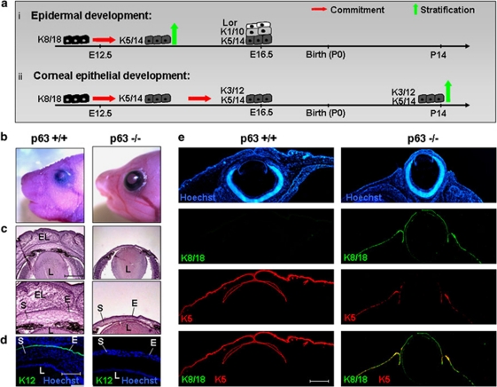
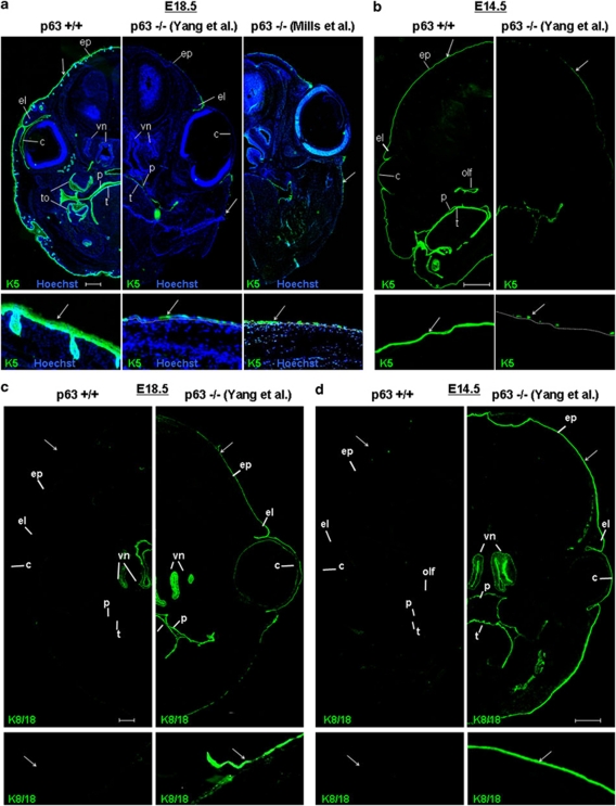
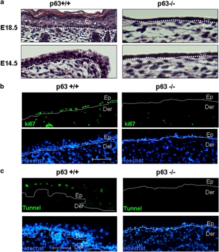
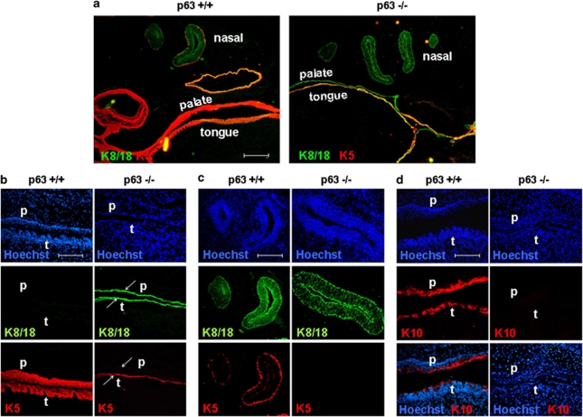
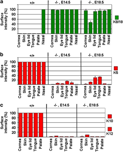
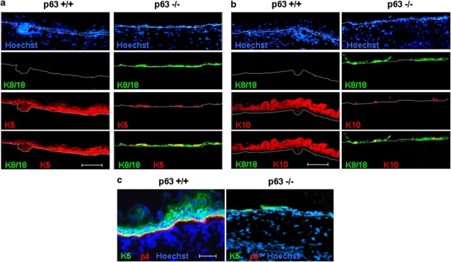
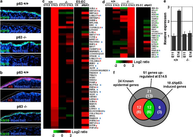
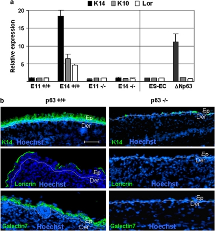
p63, a member of p53 family, has a significant role in the development and maintenance of stratified epithelia. However, a persistent dispute remained over the last decade concerning the interpretation of the severe failure of p63-null embryos to develop stratified epithelia. In this study, by investigating both p63-deficient strains, we demonstrated that p63-deficient epithelia failed to develop beyond ectodermal stage as they remained a monolayer of non-proliferating cells expressing K8/K18. Importantly, in the absence of p63, corneal-epithelial commitment (which occurs at embryonic day 12.5 of mouse embryogenesis) was hampered 3 weeks before corneal stem cell renewal (that begins at P14). Taken together, these data illustrate the significant role of p63 in epithelial embryogenesis, before and independently of other functions of p63 in adult stem cells regulation. Transcriptome analysis of laser captured-embryonic tissues confirmed the latter hypothesis, demonstrating that a battery of epidermal genes that were activated in wild-type epidermis remained silent in p63-null tissues. Furthermore, we defined a subset of novel bona fide p63-induced genes orchestrating first epidermal stratification and a subset of p63-repressed mesodermal-specific genes. These data highlight the earliest recognized action of ΔNp63 in the induction epidermal morphogenesis at E11.5. In the absence of p63, a mesodermal program is activated while epidermal morphogenesis does not initiate.


Figures

Figure 1
Figure 1
Analysis of corneal development of WT and p63−/− mice. (a) Schematic representation of epidermal (i) and corneal-epithelial (ii) development. The commitment of the ectoderm into epidermal (a(i)) and corneal (a(ii)) lineages occurs at embryonic day 12.5 of mouse embryogenesis and is hallmarked by the substitution of cytokeratins K8/K18 by K5/K14. Epidermal stratification directly follows and pluristratified epidermis is present within few days, hallmarked by typical markers, namely, K5/K14, K1/K10 and Loricrin (a(i)). However, the cornea remains static non-proliferative mono layer for 3 weeks until corneal stratification begins 2 weeks after birth (P14) on eyelids opening (a(ii)). The heads of WT (p63+/+) and the p63-deficient (p63−/−) mice (of C/BALB/c strain) were pictured (b) or processed for hematoxilin and eosin staining (c) or immunofluorescent staining for the indicated markers (d and e) at E18.5. Higher magnification of H&E staining is shown in the lower panels (c). Each panel in e contains three channels of the same field and a merge of the green and red costaining. Similar results were found in 129sv/C strain (Supplementary Figure S1). The dashed line indicates the dermal-epidermal junction. Scale bar for c and e is 500 μm, and for d is 250 μm. EL, eyelids; L, lens S, stroma; E, corneal epithelium
Figure 2
Figure 2
Mosaic images of K5 and K8/K18 immunostaining at E18.5 and E14.5. Transversal head sections of WT (p63+/+) and the indicated p63-deficient (p63−/−) mice strains were prepared for immunofluorescent staining at embryonic day 18.5 (E18.5) (a and c) or E14.5 (b and d) using anti K5 antibodies (a and b) or K8K18 antibodies (c and d). The specific p63-deficient strains are indicated in brackets. Lower panels are enlarged regions of the epidermis indicated by the arrows in the upper panels. ep, epidermis; t, tongue; p, palate; el, eyelids; c, cornea; olf, olfactory; vn, vumeronasal. Scale bars for a and b are 500 μm. Comparable observations were found in both p63−/− strains (129sv and C/BALB/c strains)
Figure 3
Figure 3
Examination of proliferation and apoptosis during first epidermal stratification. (a) Paraffin sections of the indicated embryonic skin (of C/BALB/c strain) that were prepared at E18.5 and E14.5 were used for hematoxilin and eosin staining. Tissue sections of embryonic skin (of C/BALB/c strain at E15.5) were subjected to immunostaining using ki67 antibody (b) or alternatively used for Tunel assay (c). The dashed line indicates the dermal-epidermal junction. Der, dermis; Ep, epidermis. Scale bars for b and c are 100 μm
Figure 4
Figure 4
Immunostaining of oral epithelia. Oral tissues (E18.5, C/BALB/c mouse strain) were subjected to costaining of the indicated proteins. Merges shown in a, while b and c are higher magnification of selected regions shown in a. Comparable observations were found in 129sv strains (not shown). Scale bar for a is 300 and 100 μm for bd. p, palate; t, tongue
Figure 5
Figure 5
Quantification of the immunofluorescent staining. Computerized analysis of the immunostaining shown in Figures 1, 2 and 3. The surface-intensity values of immunofluorescence staining were determined using computerized analysis as described in Materials and Methods. The relative expression was normalized as a percentage of values recorded for WT (b and c) or p63−/− (a)
Figure 6
Figure 6
Analysis of epidermal markers in embryonic epidermis. The epidermis of the indicated p63 genotypes (of C/BALB/c strain) at E18.5 was subjected to immunofluorescent costaining of the indicated proteins (ac). Each panel contains three channels of the same field and a merge of the green and red costaining is shown (a and b). The dashed line indicates the dermal-epidermal junction. Scale bars for a and b are 100 and 50 μm for c. Comparable observations were found in 129sv strain
Figure 7
Figure 7
Transcriptome analyses of embryonic epidermal tissues and of ES-derived ectodermal-like cells transfected with ΔNp63. The indicated genotypes (of C/BALB/c strain) at E11.5 (a) and E14.5 (b) were immunostained as indicated before laser capture microdissection of the epidermis (lower panels are the same fields following epidermal laser capture). Tissues were used for transcriptome analysis (c and d, left panel). In addition, murine ES-derived ectodermal cells (ES-EC) transfected with empty vector (Ctl) or ΔNp63α (ΔNp63), were subjected to transcriptome analysis (c and d, right panel). The top-rank 51 genes with highest WT/KO ratio at E14.5 are presented by heat map presentations (c, left panel). The top-rank 24 genes with highest KO/WT expression ratio at E14.5 are presented by heat map presentations (d, left panels). The corresponding expression in the transcriptome of ES-EC transfectants is presented (c and d, right panels). The expression (Log2) levels are shown (see color code below). Known epidermal or mesodermal genes are annotated by red or green asterisks in c and d, respectively. Genes that were found to contain p63-binding site are annotated by blue asterisks (c and d). The average expression of the 24 epidermal genes is presented in e. (f) Venn diagram categorizing the 51 genes that were upregulated in WT at E14.5 (gray), among them known epidermal genes (red), ΔNp63-induced genes (blue) or both (green) (e). The number of genes that also contained p63-binding site in each group is indicated in brackets (e). Scale bars for a and b are 100 μm
Figure 8
Figure 8
Validation of transcriptome analyses by qPCR and immunofluorescent staining. (a) Real-time PCR analysis was performed to amplify the indicated transcripts using the same RNA samples that were used for the micro array assay that is described in Figure 6. (b) Epidermal tissues of the indicated genotypes (of C/BALB/c strain) were immunostained for K14, Loricrin and Galectin7. The dashed line indicates the dermal-epidermal junction. Scale bar for b is 100 μm

References

    1. Blanpain C, Fuchs E. Epidermal homeostasis: a balancing act of stem cells in the skin. Nat Rev Mol Cell Biol. 2009;10:207–217. - PMC - PubMed
    1. Candi E, Cipollone R, Rivetti di Val Cervo P, Gonfloni S, Melino G, Knight R. p63 in epithelial development. Cell Mol Life Sci. 2008;65:3126–3133. - PMC - PubMed
    1. Crum CP, McKeon FD. p63 in epithelial survival, germ cell surveillance, and neoplasia. Annu Rev Pathol. 2010;5:349–371. - PubMed
    1. Fuchs E. Scratching the surface of skin development. Nature. 2007;445:834–842. - PMC - PubMed
    1. Mills AA, Zheng B, Wang XJ, Vogel H, Roop DR, Bradley A. p63 is a p53 homologue required for limb and epidermal morphogenesis. Nature. 1999;398:708–713. - PubMed

Publication types

MeSH terms