. 2011 Jan;461(1):29-44.
doi: 10.1007/s00424-010-0906-1. Epub 2010 Dec 3.

Delayed conduction and its implications in murine Scn5a(+/-) hearts: independent and interacting effects of genotype, age, and sex

Affiliations

Delayed conduction and its implications in murine Scn5a(+/-) hearts: independent and interacting effects of genotype, age, and sex

Kamalan Jeevaratnam et al. Pflugers Arch. 2011 Jan.

Abstract

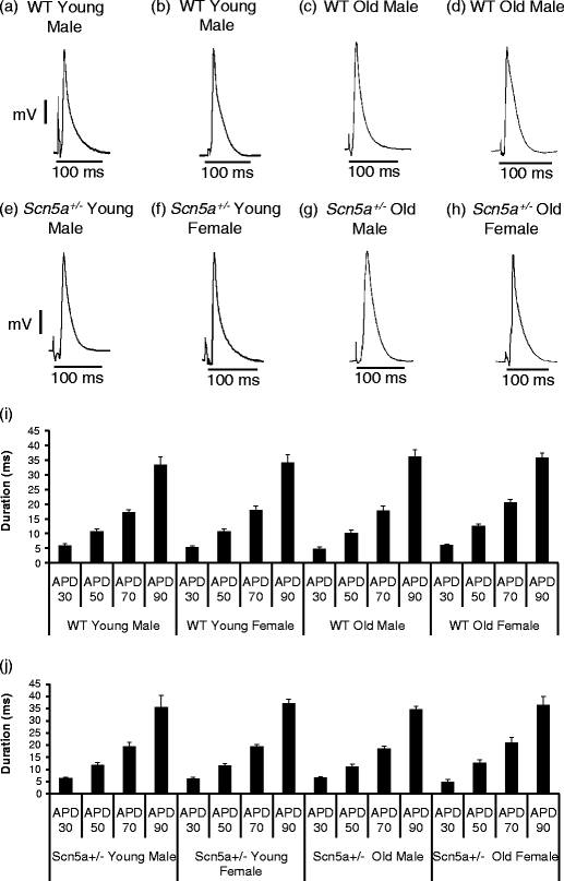
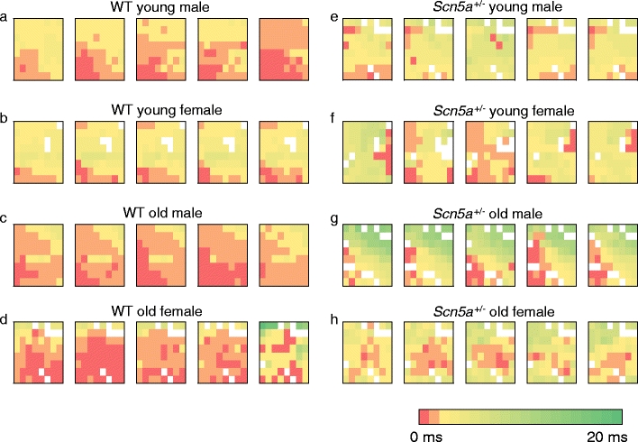
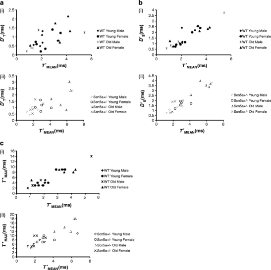
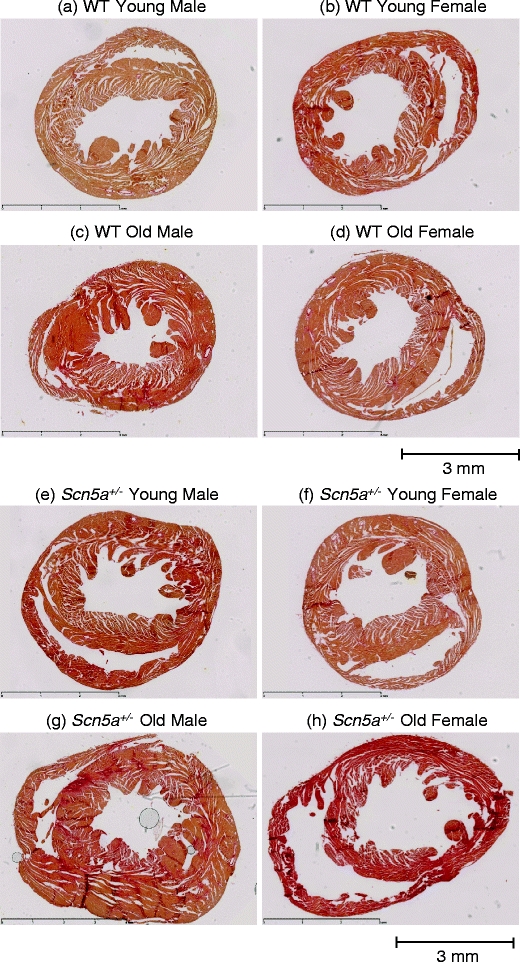
We explored for relationships between SCN5A haploinsufficiency, implicated in clinical arrhythmogenicity, and right ventricular (RV) conduction disorders in Langendorff-perfused, male and female, and young (3 months) and old (>12 month old) Scn5a ( +/-) and wild type (WT) hearts. The investigated conditions of genotype, age, and sex affected latencies but not repolarization time courses of RV monophasic action potentials. This prompted examination of the patterns of RV epicardial activation, its dispersion, and their interrelationships as possible arrhythmic mechanisms using a 64-channel, multi-electrode array. Mean ventricular activation times (T*(MEAN)), spatial dispersions (D* (S)) between recording channels/cardiac cycle, and maximum activation times (T* (MAX)) representing the slowest possible conduction in any given heart were all higher in old male Scn5a ( +/-) compared with young male and old female Scn5a ( +/-) and old male WT. Temporal dispersions (D*(T)) of recording channels were similarly higher in old male Scn5a (+/-) compared with old male WT. All groupings of D*(T), D*(S), and T*(MAX) nevertheless linearly correlated with T*(MEAN), with indistinguishable slopes. The variates explored thus influence D*(T), D*(S), and T*(MAX) through actions on T*(MEAN). These findings in turn correlated with increased levels of fibrosis in young male, young female, and old male Scn5a ( +/-) compared with the corresponding WTs. We thus demonstrate for the first time independent and interacting effects of genotype, age, and sex on epicardial conduction and its dispersions at least partially attributable to fibrotic change, resulting in the greatest effects in old male Scn5a ( +/-) in an absence of alterations in repolarization time courses. This directly implicates altered depolarization in the clinical arrhythmogenicity associated with Scn5a ( +/-).


Figures

Fig. 1
Fig. 1
Mean repolarization durations represented by values of APD30, APD50, APD70, and APD90 for WT and Scn5a+/− hearts sorted by age and sex. APDs were obtained from Langendorff-perfused hearts isolated from healthy mice. Representative MAPs waveform recordings are shown for all the eight experimental groups (ah). Durations of APDs for the stratification by genotype only, genotype and age, and genotype and sex are not shown. The full stratification of both WT and Scn5a+/− mice by both age and sex is represented by i and j, respectively. All APD values were not significantly different irrespective of genotype, age, or sex through all stratification levels
Fig. 2
Fig. 2
Representative biphasic waveform recordings. Representative recordings of 16 channels obtained from a 64-channel multi-electrode array from: WT old male (a), Scn5a +/− old male (b), WT old female (c), and Scn5a +/− old female (d). Most recording channels showed a presence of electrical activity during measurements from intrinsically beating Langendorff-perfused hearts
Fig. 3
Fig. 3
Representative activation maps of five successive cardiac cycles in WT and Scn5a +/− hearts grouped by age and sex. The study population was stratified into: WT young male (a), WT young female (b), WT old male (c), WT old female (d), Scn5a +/− young male (e), Scn5a +/− young female (f), Scn5a +/− old male (g), and Scn5a +/− old female (h). Recordings using a 64-channel multi-electrode array on intrinsically beating Langendorff-perfused mouse hearts were performed. Activation times for 64 channels were determined in five successive cardiac cycles in all hearts to produce the respective activation maps. Color gradients represent the various activation time for every channel. Time of first activation is represented as red followed by gradual color change to green representing time of last activation
Fig. 4
Fig. 4
Representative plots summarizing linear correlation and regression analysis of D*T, D*S, and T*MAX against T*MEAN. Plots for D*T (a), D*S (b), and T*MAX (c) for WT (i) and Scn5a +/− (ii). The regression analysis demonstrated significant correlations between the parameters of D*T, D*S, and T*MAX on the ordinate and T*MEAN on the abscissa with slopes that were not significantly different whether following stratification by genotype, age, or sex
Fig. 5
Fig. 5
Representative slide for cardiac fibrosis staining in WT and Scn5a +/− hearts grouped by age and sex. The study population was stratified into: WT young male (a), WT young female (b), WT old male (c), WT old female (d), Scn5a +/− young male (e), Scn5a +/− young female (f), Scn5a +/− old male (g), and Scn5a +/− old female (h). Hearts were routinely stained with Sirius red, and morphometric analysis for percentage of fibrosis was performed for all eight groups. Areas of increased red uptake signify presence of fibrotic changes. Horizontal bar below sections in each panel denotes a 3mm distance

References

    1. Alings M, Wilde A. “Brugada” syndrome: clinical data and suggested pathophysiological mechanism. Circulation. 1999;99:666–673. - PubMed
    1. Antzelevitch C. Brugada syndrome: historical perspectives and observations. Eur Heart J. 2002;23:676–678. doi: 10.1053/euhj.2001.3145. - DOI - PubMed
    1. Caldwell BJ, Trew ML, Sands GB, Hooks DA, LeGrice IJ, Smaill BH. Three distinct directions of intramural activation reveal nonuniform side-to-side electrical coupling of ventricular myocytes. Circ Arrhythm Electrophysiol. 2009;2:433–440. doi: 10.1161/CIRCEP.108.830133. - DOI - PubMed
    1. Capulzini L, Brugada P, Brugada J, Brugada R. Arrhythmia and right heart disease: from genetic basis to clinical practice. Rev Esp Cardiol. 2010;63:963–983. - PubMed
    1. Gussak I, Antzelevitch C, Bjerregaard P, Towbin JA, Chaitman BR. The Brugada syndrome: clinical, electrophysiologic and genetic aspects. J Am Coll Cardiol. 1999;33:5–15. doi: 10.1016/S0735-1097(98)00528-2. - DOI - PubMed

Publication types

Substances

LinkOut - more resources