

Molecular diagnostics in embryonal brain tumors
- PMID: 21129063
- PMCID: PMC3575597
- DOI: 10.1111/j.1750-3639.2010.00455.x
Molecular diagnostics in embryonal brain tumors
Abstract
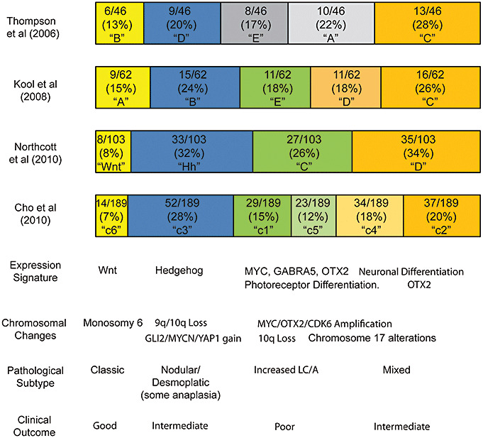
Embryonal brain tumors are a heterogeneous group of neoplasms united by the presence of poorly differentiated stem-like cells. Molecular details are increasingly being used to separate them into biologically and clinically meaningful groups. For medulloblastoma, integrated mRNA expression profiling and DNA analysis by a number of research groups defines 4-6 distinctive molecular variants. A subset with prominent Wnt activity is associated with good clinical outcomes and classic histology. Medulloblastomas showing a Hedgehog gene expression signature are frequently of the desmoplastic/nodular subtype. Interestingly, Hedgehog activity is found in tumors arising either in infants or older teenagers and adults. The association of clinically aggressive medulloblastoma with MYC expression, large cell/anaplastic change and high levels of photoreceptor differentiation transcripts has also been noted in several studies. Immunohistochemical analysis of just one or two genes per molecular medulloblastoma variant may be sufficient for accurate classification, and this would be of great practical utility if validated. Advances have also been made in the classification of central nervous system (CNS) Primitive Neuroectodermal Tumors (PNET), as several groups have identified an amplicon at chromosome 19q13.41-42, which appears to define a unique PNET subtype associated with prominent true rosettes, young age and very poor outcomes.
© 2010 The Author; Brain Pathology © 2010 International Society of Neuropathology.
Figures
References
-
- Aldosari N, Bigner SH, Burger PC, Becker L, Kepner JL, Friedman HS, McLendon RE (2002) MYCC and MYCN oncogene amplification in medulloblastoma. A fluorescence in situ hybridization study on paraffin sections from the Children's Oncology Group. Arch Pathol Lab Med 126:540–544. - PubMed
-
- Badiali M, Pession A, Basso G, Andreini L, Rigobello L, Galassi E, Giangaspero F (1991) N‐myc and c‐myc oncogenes amplification in medulloblastomas. Evidence of particularly aggressive behavior of a tumor with c‐myc amplification. Tumori 77:118–121. - PubMed
-
- Baeza N, Masuoka J, Kleihues P, Ohgaki H (2003) AXIN1 mutations but not deletions in cerebellar medulloblastomas. Oncogene 22:632–636. - PubMed
-
- Batra SK, McLendon RE, Koo JS, Castelino‐Prabhu S, Fuchs HE, Krischer JP et al (1995) Prognostic implications of chromosome 17p deletions in human medulloblastomas. J Neurooncol 24:39–45. - PubMed
Publication types
MeSH terms
Grants and funding
LinkOut - more resources
Full Text Sources
Medical
