. 2011 Apr;13(4):602-19.
doi: 10.1111/j.1462-5822.2010.01557.x. Epub 2010 Dec 28.

Alix regulates egress of hepatitis B virus naked capsid particles in an ESCRT-independent manner

Affiliations

Alix regulates egress of hepatitis B virus naked capsid particles in an ESCRT-independent manner

Andreas Bardens et al. Cell Microbiol. 2011 Apr.

Abstract

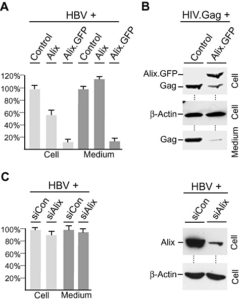
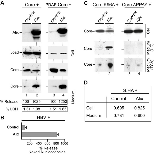
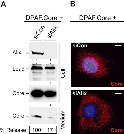
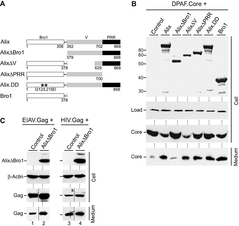
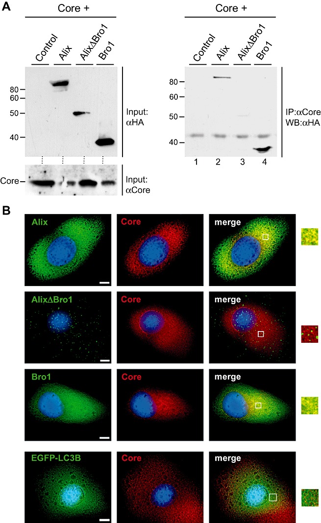
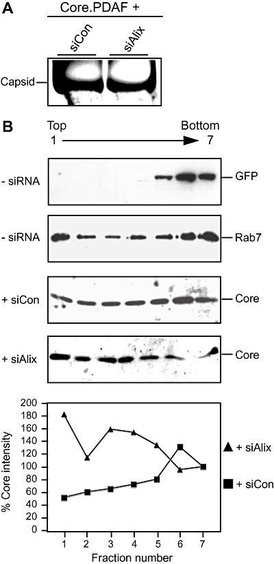
Hepatitis B virus (HBV) is an enveloped DNA virus that exploits the endosomal sorting complexes required for transport (ESCRT) pathway for budding. In addition to infectious particles, HBV-replicating cells release non-enveloped (nucleo)capsids, but their functional implication and pathways of release are unclear. Here, we focused on the molecular mechanisms and found that the sole expression of the HBV core protein is sufficient for capsid release. Unexpectedly, released capsids are devoid of a detectable membrane bilayer, implicating a non-vesicular exocytosis process. Unlike virions, naked capsid budding does not require the ESCRT machinery. Rather, we identified Alix, a multifunctional protein with key roles in membrane biology, as a regulator of capsid budding. Ectopic overexpression of Alix enhanced capsid egress, while its depletion inhibited capsid release. Notably, the loss of Alix did not impair HBV production, furthermore indicating that virions and capsids use diverse export routes. By mapping of Alix domains responsible for its capsid release-mediating activity, its Bro1 domain was found to be required and sufficient. Alix binds to core via its Bro1 domain and retained its activity even if its ESCRT-III binding site is disrupted. Together, the boomerang-shaped Bro1 domain of Alix appears to escort capsids without ESCRT.


Figures

Figure 1
Figure 1
HBV naked capsid release from cells. A. HuH‐7 cells were transfected with empty plasmid DNA (Control), the HBV replicon under foreign promoter control (pHBV), the untagged core construct (pCore) or the DPAF‐tagged core construct (pDPAF.Core). Three days after transfection, lysates were prepared with NP‐40 and immunoblotted with a polyclonal core antiserum (K46). A non‐specific band stained by the antiserum serves as a control for identical gel loading (Load). Capsids released into the medium were recovered by ultracentrifugation and analysed by non‐denaturing dot‐blot analysis using a capsid‐specific antibody (3HB17). To probe for cell cytotoxicity, supernatants were assessed for LDH activity. B. Sensitivity analysis of the LDH assay. Supernatants of control‐, pCore‐ or VSV‐G‐transfected cells were assayed for LDH, and the transfection rates were determined by immunofluorescence analysis using core‐ and VSV‐G‐specific antibodies. LDH levels are shown in % amount relative to 3 × 103 cells expressing the constructs. C. Released core particles resemble authentic capsids. Concentrated culture medium of pCore‐transfected cells was applied to isopycnic caesium chloride gradient centrifugation and fractions were assayed by a core‐specific (HBcAg) ELISA. D and E. Released capsids do not contain a lipid envelope. (D) Concentrated culture medium of control‐ or core‐transfected cells were mock‐treated or treated with NP‐40 prior to the capsid‐specific dot‐blot analysis. (E) Extracellular capsids were left untreated or treated with proteinase K (PK) in the absence or presence of NP‐40, and samples were analysed by core‐specific immunoblotting (K46).
Figure 2
Figure 2
HBV naked capsid release does not require ESCRT functions. A. ESCRT‐I‐independent capsid budding. HuH‐7 cells were treated with siRNAs targeting Tsg101, Vps28 or control siRNA. Two days later, cells were transfected with the DPAF.Core construct. After an additional 2 days, cells were lysed with SDS buffer and extracellular capsids were recovered by ultracentrifugation. Lysates were subjected to Tsg101‐ and Vps28‐specific Western blotting to demonstrate depletion. For core detection, lysates and supernatants were analysed by anti‐DPAF immunoblotting. To confirm equal loading of cell lysates, we took advantage that the anti‐DPAF antibody specifically cross‐reacts with an endogenous protein of HuH‐7 cells of unknown nature (Load). B and C. ESCRT‐III‐ and Vps4‐independent capsid budding. DPAF.Core was transfected into HuH‐7 cells with either empty plasmid DNA (Control) or WT or DN versions of CHMP3 and Vps4A, as indicated above each lane. Co‐transfections were performed, respectively, at a 3:1 DNA ratio. Three days post transfection, SDS lysates were prepared and analysed by FLAG‐ or GFP‐specific Western blotting to demonstrate expression of CHMP3.wt or CHMP3.dn, Vps4A.wt and Vps4A.dn respectively. Cell‐associated core and capsids released into the media were probed by anti‐DPAF immunoblotting. D. HuH‐7 cells were transfected with the core expression vector (Core) or the HBV replicon (HBV) and immunostained with rabbit anti‐capsid (K45) and mouse anti‐LBPA antibodies. After staining with AlexaFluor 546‐conjugated anti‐rabbit and AlexaFluor 488‐conjugated anti‐mouse antibodies, cells were visualized by deconvolution fluorescence microscopy. The overlays of the fluorescences are shown in the right column with yellow colour indicating colocalization. DNA staining of the nuclei is in blue. Bar, 10 µm.
Figure 3
Figure 3
HBV budding requires ESCRT, but not Alix. A. DN Alix blocks HBV budding. HuH‐7 cells were co‐transfected with the HBV replicon and empty plasmid DNA (Control), HA‐tagged WT Alix or the GFP‐tagged Alix mutant (Alix.GFP) at a 1:3 DNA weight ratio respectively. Three days post transfection, cellular supernatants (Medium; black columns) and cytoplasmic extracts prepared with Triton X‐100 (Cell; grey columns) were harvested. HBV release was detected by envelope‐specific immunoprecipitation of supernatants and real‐time PCR of the viral genomes. Non‐enveloped cytoplasmic nucleocapsids were immunoprecipitated with anti‐capsid antibodies (K45) and analysed by PCR. PCR results were demonstrated in per cent amount relative to control‐transfected cells. B. DN Alix blocks HIV.Gag budding. GFP‐tagged HIV.Gag was co‐transfected with control DNA or the Alix.GFP construct at a 1:3 ratio. NP‐40 lysates and VLPs harvested from the supernatants were analysed by GFP‐ and β‐actin‐specific immunoblotting. C. Alix depletion does not inhibit HBV budding. HuH‐7 cells were transfected with control siRNA or siRNA against Alix. Two days later, cells were retransfected with the HBV replicon and harvested after additional 3 days. Intracellular nucleocapsids and extracellular virions were assayed as in (A). Mean results of four PCR reactions are demonstrated in per cent amount relative to control‐treated cells (left). To probe for Alix depletion, the same lysates were immunoblotted with anti‐Alix and anti‐β‐actin antibodies (right).
Figure 4
Figure 4
Excess Alix enhances HBV naked (nucleo)capsid release. A. Core (lanes 1 and 2) or DPAF‐tagged core (lanes 3 and 4) was co‐transfected with control DNA or HA‐tagged Alix at a 1:3 DNA ratio respectively. Cell extracts were prepared with SDS and analysed by HA‐specific immunoblotting to monitor expression of Alix. For detection of core, the core antiserum (K46) (lanes 1 and 2) or the DPAF antibody (lanes 3 and 4) was used. In either case, non‐specifically stained bands served as a control for gel loading (Load). Capsids harvested from the culture media were analysed in the same manner. The experiments were repeated three times, and capsids released into the supernatants were quantified and demonstrated in per cent amount relative to control cells. To probe for cell lysis, supernatants were assayed for LDH activity. B. The pHBV replicon was co‐transfected with Alix or control plasmid DNA. Cell supernatants were immunoprecipitated with the capsid‐specific antiserum (K45) prior to PCR measurement of the viral DNA. Mean PCR results are demonstrated in per cent amount relative to control‐transfected cells. C. The core mutants Core.K96A (lanes 1 and 2) and CoreΔPPAY (lanes 3 and 4) were subjected to the co‐transfection assay exactly as in (A). Cell lysates and media concentrated by either ultracentrifugation (UC) or TCA precipitation (TCA) were immunoblotted with the core antiserum (K46). D. An HA‐tagged version of the HBV small envelope protein (S.HA) was transfected into HuH‐7 cells together with Alix or a control construct at a 1:3 ratio respectively. Amounts of S were examined by ELISA and are expressed as mean units of optical density at 492 nm (n = 3).
Figure 5
Figure 5
Alix is essential for HBV naked capsid release. A. HuH‐7 cells treated with control‐ or Alix‐specific siRNAs were retransfected with DPAF.Core. SDS lysates and concentrated supernatants were Western blotted with anti‐Alix and anti‐DPAF antibodies. A non‐specifically stained band served as a control for gel loading (Load). Capsids released into the supernatants were quantified and demonstrated in per cent amount relative to control‐depleted cells. B. Loss of Alix affects the intracellular distribution of core. Cells were transfected as in (A) and immunostained with anti‐capsid antibodies (K45) followed by staining with AlexaFluor 546‐conjugated anti‐rabbit antibodies. DNA staining of the nuclei is in blue. Bar, 10 µm.
Figure 6
Figure 6
Mapping of Alix domains responsible for its HBV capsid release‐mediating activity. A. Schematic representation of WT Alix and its mutants. The domain architecture of Alix with the Bro1, V and PRD domains is depicted. Numbers below refer to aa positions, and the two stars in Alix.DD denote point mutations introduced at aa positions 212 and 216. B. DPAF.Core was co‐transfected with HA‐tagged WT or the indicated Alix mutants at a 1:3 ratio. Following transient expression, cells were lysed with SDS and supernatants were subjected to ultracentrifugation. Synthesis of wt Alix and its mutants is shown by HA‐specific immunoblotting. Protein levels of intracellular cores and extracellular capsids were examined by DPAF‐specific Western blotting. Uniformity of sample loading is shown by a band cross‐reacting with the DPAF antibody (Load). Numbers to the left of the top panel refer to molecular weight standards in kDa. C. EGFP‐tagged EIAV.Gag or HIV.Gag were co‐transfected with control DNA or HA‐tagged AlixΔBro1 at a 1:3 DNA ratio respectively. Gag expression and VLP release efficiency were analysed by GFP‐specific immunoblotting, while synthesis of AlixΔBro1 was determined by anti‐HA Western blotting.
Figure 7
Figure 7
HBV core interacts and colocalizes with the Bro1 domain of Alix. A. HuH‐7 cells were co‐transfected with core together with HA‐tagged wt or mutant Alix, as denoted above each lane. Each co‐transfection was performed at a 3:1 DNA ratio, respectively, and empty plasmid was used as a negative control. Synthesis (Input) of core and the Alix constructs is shown by immunoblotting of lysates with anti‐core (K46) and anti‐HA antibodies. Input amounts correspond to 10% of the samples used for immune capture (left). For co‐immunoprecipitation, lysates were incubated with anti‐capsid antibodies (K45) before Western blotting (WB) with the HA‐specific antibody (right). B. Core colocalizes with Alix and Bro1, but not with AlixΔBro1. Cells were co‐transfected with core plus HA‐tagged Alix, AlixΔBro1 or Bro1 and immunostained with rabbit anti‐core (K45) and mouse anti‐HA antibodies. For control, core was co‐transfected with EGFP‐LC3B, a cytosolic marker protein. After staining with secondary antibodies, cells were analysed by deconvolution fluorescence microscopy. The staining pattern of the Alix constructs and the autofluorescence of EGFP‐LC3B are shown in green, and the fluorescent signal of core is in red. The overlays of the fluorescence patterns are shown in the right column with yellow colour indicating colocalization. DNA staining is shown in blue. Bar, 10 µm.
Figure 8
Figure 8
Alix is not required for capsid assembly and membrane association of core. A. HuH‐7 cells treated with control‐ or Alix‐specific siRNAs were retransfected with DPAF.Core. Cytoplasmic capsids were concentrated by PEG precipitation and separated in a native agarose gel, blotted and detected with anti‐core (K45). B. Cells were either left untreated or treated with control‐ or Alix‐specific siRNAs, as indicated to the left of the panels. Subsequent DNA transfections were performed with a GFP construct, GFP‐tagged Rab7 or core, as denoted on the right of the panels. Cell extracts were subjected to density flotation analyses, gradients were fractioned from the top, and fractions were analysed by GFP‐ or core‐specific immunoblotting. The graph (bottom) shows quantification of the core signals present in the gradient fractions of siCon‐ and siAlix‐treated cells. The band intensities were quantified and demonstrated in per cent amount relative to the corresponding gradient bottom fractions (fraction 7).

Similar articles

Cited by

References

    1. Arzberger, S. , Hosel, M. , and Protzer, U. (2010) Apoptosis of hepatitis B virus infected hepatocytes prevents release of infectious virus. J Virol 84: 11994–12001. - PMC - PubMed
    1. Babst, M. , Sato, T.K. , Banta, L.M. , and Emr, S.D. (1997) Endosomal transport function in yeast requires a novel AAA‐type ATPase, Vps4p. EMBO J 16: 1820–1831. - PMC - PubMed
    1. Beterams, G. , and Nassal, M. (2001) Significant interference with hepatitis B virus replication by a core–nuclease fusion protein. J Biol Chem 276: 8875–8883. - PubMed
    1. Bishop, N. , and Woodman, P. (2001) TSG101/mammalian VPS23 and mammalian VPS28 interact directly and are recruited to VPS4‐induced endosomes. J Biol Chem 276: 11735–11742. - PubMed
    1. Bruss, V. (2007) Hepatitis B virus morphogenesis. World J Gastroenterol 13: 65–73. - PMC - PubMed

Publication types

MeSH terms

Substances