. 2010 Dec 3:9:351.
doi: 10.1186/1475-2875-9-351.

Dynamic subcellular localization of isoforms of the folate pathway enzyme serine hydroxymethyltransferase (SHMT) through the erythrocytic cycle of Plasmodium falciparum

Affiliations

Dynamic subcellular localization of isoforms of the folate pathway enzyme serine hydroxymethyltransferase (SHMT) through the erythrocytic cycle of Plasmodium falciparum

Martin Read et al. Malar J. .

Abstract

Background: The folate pathway enzyme serine hydroxymethyltransferase (SHMT) converts serine to glycine and 5,10-methylenetetrahydrofolate and is essential for the acquisition of one-carbon units for subsequent transfer reactions. 5,10-methylenetetrahydrofolate is used by thymidylate synthase to convert dUMP to dTMP for DNA synthesis. In Plasmodium falciparum an enzymatically functional SHMT (PfSHMTc) and a related, apparently inactive isoform (PfSHMTm) are found, encoded by different genes. Here, patterns of localization of the two isoforms during the parasite erythrocytic cycle are investigated.

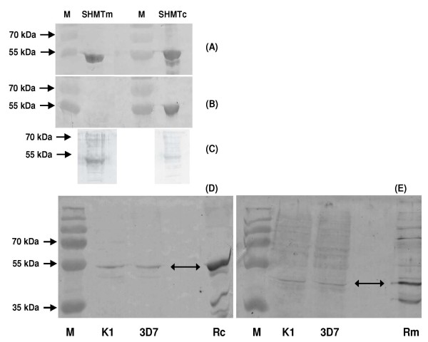
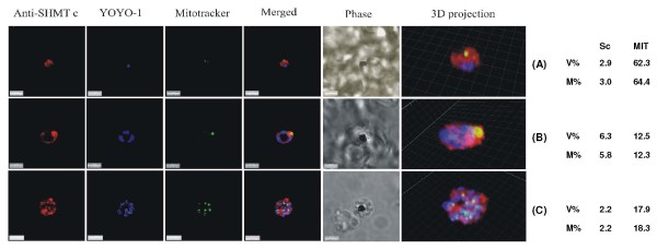
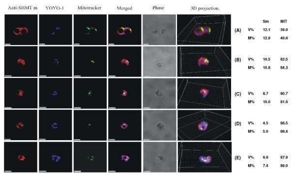
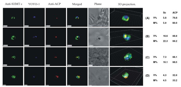
Methods: Polyclonal antibodies were raised to PfSHMTc and PfSHMTm, and, together with specific markers for the mitochondrion and apicoplast, were employed in quantitative confocal fluorescence microscopy of blood-stage parasites.

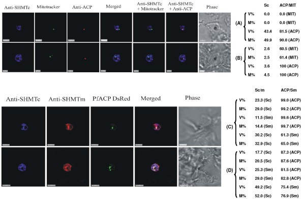
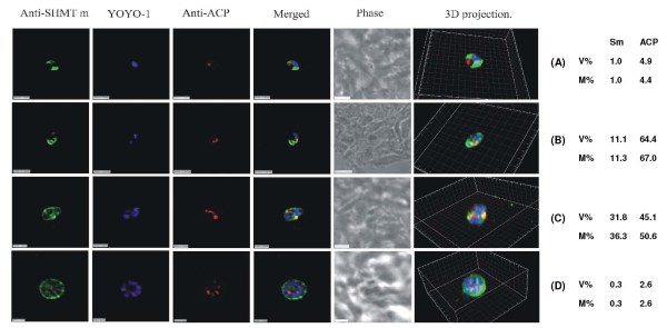
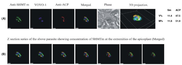
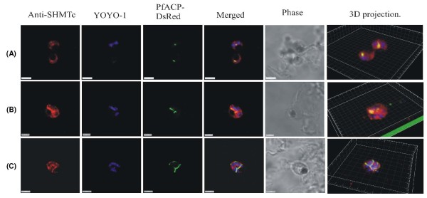
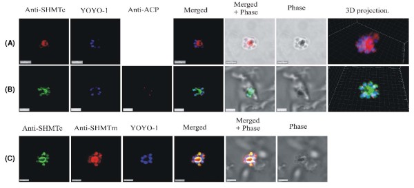
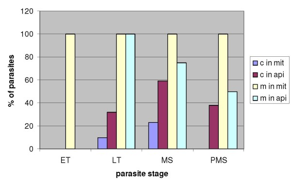
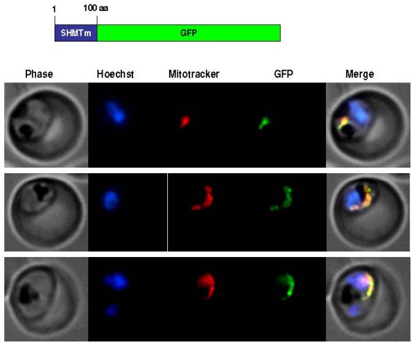
Results: As well as the expected cytoplasmic occupancy of PfSHMTc during all stages, localization into the mitochondrion and apicoplast occurred in a stage-specific manner. Although early trophozoites lacked visible organellar PfSHMTc, a significant percentage of parasites showed such fluorescence during the mid-to-late trophozoite and schizont stages. In the case of the mitochondrion, the majority of parasites in these stages at any given time showed no marked PfSHMTc fluorescence, suggesting that its occupancy of this organelle is of limited duration. PfSHMTm showed a distinctly more pronounced mitochondrial location through most of the erythrocytic cycle and GFP-tagging of its N-terminal region confirmed the predicted presence of a mitochondrial signal sequence. Within the apicoplast, a majority of mitotic schizonts showed a marked concentration of PfSHMTc, whose localization in this organelle was less restricted than for the mitochondrion and persisted from the late trophozoite to the post-mitotic stages. PfSHMTm showed a broadly similar distribution across the cycle, but with a distinctive punctate accumulation towards the ends of elongating apicoplasts. In very late post-mitotic schizonts, both PfSHMTc and PfSHMTm were concentrated in the central region of the parasite that becomes the residual body on erythrocyte lysis and merozoite release.

Conclusions: Both PfSHMTc and PfSHMTm show dynamic, stage-dependent localization among the different compartments of the parasite and sequence analysis suggests they may also reversibly associate with each other, a factor that may be critical to folate cofactor function, given the apparent lack of enzymic activity of PfSHMTm.


Figures

Figure 1
Figure 1
Specificity of the polyclonal anti-PfSHMT preparations. Full-length His-tagged recombinant protein (500 ng) expressed from the genes encoding PfSHMTc (PFL1720w) and PfSHMTm (Pf14_0534) probed on western blots with (A) anti-polyhistidine IgG, (B) the anti-PfSHMTc and (C) the anti-PfSHMTm preparations used for subsequent immunofluorescence studies. Panels D and E are western blots of total parasite extracts from K1 and 3D7 probed with anti-PfSHMTc (D) and anti-PfSHMTm (E). Rc, recombinant PfSHMTc; Rm, recombinant PfSHMTm; M, prestained molecular weight markers.
Figure 2
Figure 2
PfSHMTc immunofluorescence images showing localization in the mitochondrion. (A) Mid-trophozoite showing the association of a small mitochondrion with PfSHMTc fluorescence. (B) Early schizont showing association of an enlarged globular mitochondrion with a region of more intense PfSHMTc fluorescence. (C) Late schizont showing very little co-localization of mitochondria with areas of PfSHMTc fluorescence. Mitochondria are closely aligned to nuclei and show some co-localization with YOYO1 staining (scale bars 3 μm). The associated table shows the percentage volume (V%) and material (M%) co-localization data for PfSHMTc (Sc) and MitoTracker (MIT) fluorescence.
Figure 3
Figure 3
PfSHMTm immunofluorescence images showing localization in the mitochondrion. (A) Two late trophozoites. (B-D) Mitotic schizonts. (E) Post-mitotic schizont. The images show the persistence of co-localization of PfSHMTm fluorescence with the mitochondria throughout the developmental cycle (scale bars 3 μm). The associated table shows the percentage volume (V%) and material (M%) co-localization data for PfSHMTm (Sm) and MitoTracker (MIT) fluorescence.
Figure 4
Figure 4
PfSHMTc immunofluorescence images showing localization in the apicoplast. (A) Mid-trophozoite showing the co-localization of plastid specific fluorescence with PfSHMTc fluorescence. (B) Early mitotic schizont showing very marked co-localization of plastid specific fluorescence (enlarged globular apicoplast) with PfSHMTc fluorescence. (C) Mitotic schizont showing very marked co-localization of plastid specific fluorescence with PfSHMTc fluorescence. The plastid here is in the early stages of elongation. Note also the small punctate concentration of PfSHMTc fluorescence on the periphery of the unstained region of the parasite corresponding to the pigment vacuole. (D) Mitotic schizont developmentally a little later than (C) showing a mitochondrion in the early stages of ramification. The area of intense PfSHMTc fluorescence follows the 'Y' shape of the mitochondrion closely (scale bars 3 μm, except (D) which is 2 μm). The associated table shows the percentage volume (V%) and material (M%) co-localization data for PfSHMTc (Sc) and acyl carrier protein (ACP) fluorescence.
Figure 5
Figure 5
Triple-labelling experiments. (A) and (B) Combined mitochondrial and apicoplast images probed with anti-PfSHMTc. These do not show nuclear morphology, therefore the erythrocytic cycle stage cannot be precisely ascertained; however, the size of the organelles and overall size of the parasites in (A) and (B) suggest that both are mid trophozoites. In (A) the parasite is probed with anti-PfSHMTc, MitoTracker and anti-ACP (plastid). The plastid is coincident with an area of marked PfSHMTc fluorescence, whereas the mitochondrion shows no evidence of coincident PfSHMTc fluorescence. In (B) the parasite is probed with anti-PfSHMTc, MitoTracker and anti-ACP (plastid). The plastid is coincident with a discrete area of PfSHMTc fluorescence, whereas the mitochondrion is located in a pocket of lower PfSHMTc fluorescence. (C) Parasite is probably a late trophozoite and (D) a mitotic schizont. Both parasites were expressing DsRED-labelled ACP and were probed with both anti-PcSHMTc (IgY) and anti-PfSHMTm (IgG). The distribution of the two SHMT fluorescence signals are similar but not identical, and both co-localize with the apicoplast (scale bars (A) and (C), 3 μm, (B) 2 μm, (D) 4 μm). The associated table shows the percentage volume (V%) and material (M%) co-localization data for PfSHMTc (Sc), PfSHMTm (Sm), MitoTracker (MIT) and acyl carrier protein (ACP) fluorescence.
Figure 6
Figure 6
PfSHMTm immunofluorescence images showing localization in the apicoplast. (A) Early trophozoite showing no apicoplast PfSHMTm co-localization. (B) Early mitotic schizont with PfSHMTm fluorescence conforming closely to the 'C' shaped apicoplast. (C) Mitotic schizont with an elongating apicoplast, PfSHMTm fluorescence is concentrated within the distal portions of the organelle with little fluorescence in the medial section. (D) Post-mitotic schizont showing little spatial coincidence of PfSHMTm and the multiple apicoplasts (scale bars 3 μm, except (D). which is 2 μm). The associated table shows the percentage volume (V%) and material (M%) co-localization data for PfSHMTm (Sm) and acyl carrier protein (ACP) fluorescence.
Figure 7
Figure 7
PfSHMTm apicoplast immunofluorescent images illustrating the concentration of fluorescence in the extremities of elongating apicoplasts. (A) Mitotic schizont with an elongating apicoplast. (B) A z-stack series with an interval of 0.2 μm through the same parasite showing the concentration of PfSHMTm fluorescence in the distal portions of the apicoplast and the relative lack of PfSHMTm fluorescence in its medial section (scale bars 2 μm).
Figure 8
Figure 8
Positive control images using endogenously expressed DsRED-tagged ACP instead of anti-ACP antibodies. The use of only one primary antibody, anti-PfSHMTc, with expressed DsRED tagged Pf ACP, was aimed at eliminating any possibility of artifactual fluorescence arising from interactions between two primary antibodies used simultaneously. (A) Two parasites, upper parasite is undergoing its first division, lower parasite is a late trophozoite. (B) Mitotic schizont with elongating apicoplast. (C) Mitotic schizont with ramifying apicoplast. All parasites show co-localization of anti-PfSHMTc fluorescence with the apicoplast, closely following the shape of the organelle, identical results to those obtained using two primary antibodies (scale bars (A) and (C) 3 μm, (B) 2 μm).
Figure 9
Figure 9
Late schizonts show a central concentration of PfSHMTc fluorescence. (A) Post-mitotic schizont showing a concentration of PfSHMTc fluorescence in the centre of the parasite, and overlapping the outer zone of haemozoin. PfSHMTc is largely excluded from the nuclei. (B) Post-mitotic schizont showing a concentration of PfSHMTc fluorescence in the centre of the parasite as well as at low intensity in the multiple small apicoplasts. Note the merozoite buds arranged in a radial pattern centred on the future residual body. (C) A post-mitotic parasite probed with both anti-PfSHMTc (IgY) and anti-PfSHMTm (IgG). Both SHMT proteins show a similar, but not identical distribution, as described for image series (A) and (B) above (scale bars 3 μm).
Figure 10
Figure 10
Organellar distribution of fluorescence through the erythrocytic cycle. Percentages of parasites (of the total number scanned for each stage) showing marked fluorescence for PfSHMTc (c) and PfSHMTm (m) in the mitochondrion (mit) and apicoplast (api). ET, early trophozoites; LT, late trophozoites; MS, mitotic schizonts; PMS, post-mitotic schizonts. For organellar localization of PfSHMTc, n = 82; for that of PfSHMTm, n = 76.
Figure 11
Figure 11
GFP-tagging of truncated PfSHMTm in transfected 3D7 parasites. Fluorescence images of parasites transfected to yield a GFP-fusion carrying the first 100 amino acids of PfSHMTm at the N-terminus. MitoTracker was also used to localize the mitochondrion, which showed complete coincidence with the GFP fluorescence (three examples shown).
Figure 12
Figure 12
Potential interactions between PfSHMTc and PfSHMTm. The two protein sequence motifs identified in mammalian SHMTs (both cytoplasmic and mitochondrial isoforms; top two lines) essential for stable tetramer formation; bacterial SHMTs lack these motifs entirely and form stable dimers (bottom line; E. coli as example) [42]. SHMTc from P. falciparum and its orthologues from other Plasmodium species (P. vivax, P. knowlesi, P. yoelii and P. berghei, respectively) possess a highly conserved equivalent of Motif 1 only (blue text), whereas the SHMTm forms possess only Motif 2 (red text), suggesting that heterotetramers could be formed by a combination of (PfSHMTc)2 and (PfSHMTm)2.

Similar articles

Cited by

References

    1. Snow RW, Guerra CA, Noor AM, Myint HY, Hay SI. The global distribution of clinical episodes of Plasmodium falciparum malaria. Nature. 2005;434:214–217. doi: 10.1038/nature03342. - DOI - PMC - PubMed
    1. Hyde JE. Drug-resistant malaria - an insight. FEBS Journal. 2007;274:4688–4698. doi: 10.1111/j.1742-4658.2007.05999.x. - DOI - PMC - PubMed
    1. Müller IB, Hyde JE. Antimalarial drugs: modes of action and mechanisms of parasite resistance. Future Microbiology. 2010;5:1857–1875. - PubMed
    1. Hyde JE. Exploring the folate pathway in Plasmodium falciparum. Acta Trop. 2005;94:191–206. - PMC - PubMed
    1. Krungkrai J, Webster HK, Yuthavong Y. De novo and salvage biosynthesis of pteroylpentaglutamates in the human malaria parasite, Plasmodium falciparum. Mol Biochem Parasitol. 1989;32:25–37. doi: 10.1016/0166-6851(89)90126-6. - DOI - PubMed

Publication types

MeSH terms

Substances

LinkOut - more resources