Review
. 2010 Dec 3:7:357.
doi: 10.1186/1743-422X-7-357.

Assembly and dynamics of the bacteriophage T4 homologous recombination machinery

Affiliations
Review

Assembly and dynamics of the bacteriophage T4 homologous recombination machinery

Jie Liu et al. Virol J. .

Abstract

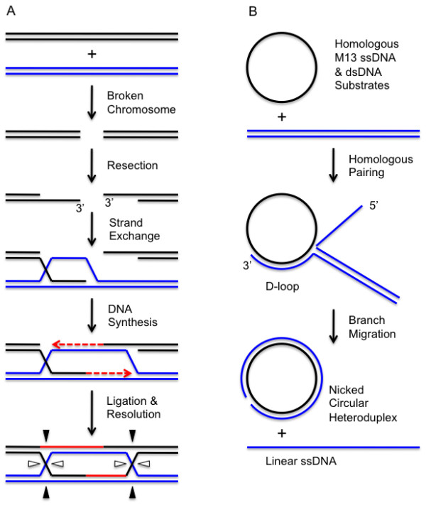
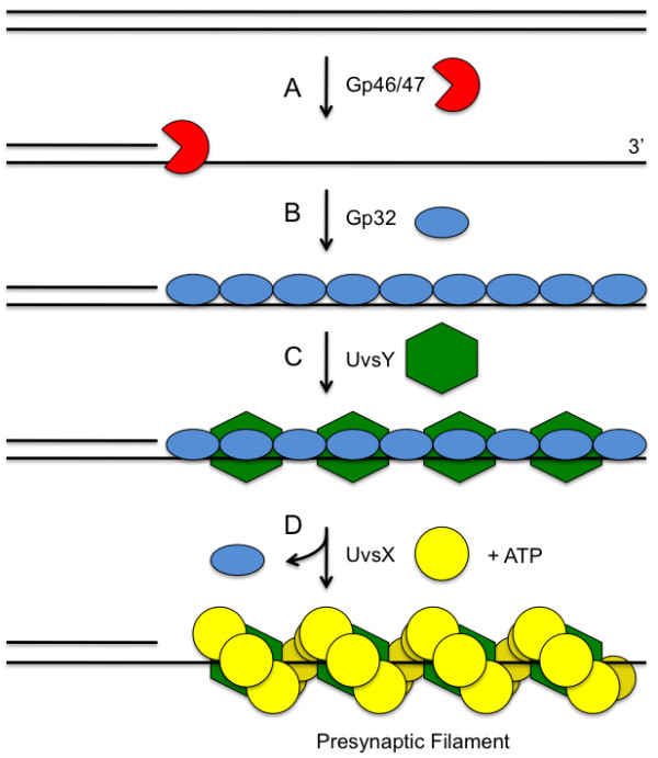
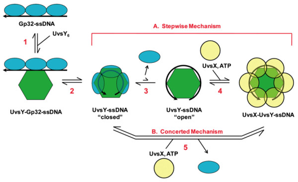
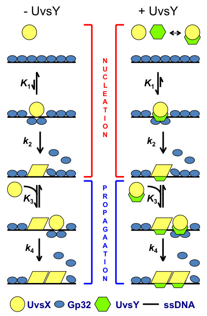
Homologous recombination (HR), a process involving the physical exchange of strands between homologous or nearly homologous DNA molecules, is critical for maintaining the genetic diversity and genome stability of species. Bacteriophage T4 is one of the classic systems for studies of homologous recombination. T4 uses HR for high-frequency genetic exchanges, for homology-directed DNA repair (HDR) processes including DNA double-strand break repair, and for the initiation of DNA replication (RDR). T4 recombination proteins are expressed at high levels during T4 infection in E. coli, and share strong sequence, structural, and/or functional conservation with their counterparts in cellular organisms. Biochemical studies of T4 recombination have provided key insights on DNA strand exchange mechanisms, on the structure and function of recombination proteins, and on the coordination of recombination and DNA synthesis activities during RDR and HDR. Recent years have seen the development of detailed biochemical models for the assembly and dynamics of presynaptic filaments in the T4 recombination system, for the atomic structure of T4 UvsX recombinase, and for the roles of DNA helicases in T4 recombination. The goal of this chapter is to review these recent advances and their implications for HR and HDR mechanisms in all organisms.


Figures

Figure 1
Figure 1
DNA strand exchange assay and the role of DNA strand exchange in double-strand break repair. Chromosome breakage is followed by nucleolytic resection to generate 3' ssDNA tails on the broken ends. The exposed ssDNA tails are the substrates for DNA strand exchange catalyzed by recombinases of the RecA/Rad51/UvsX family in collaboration with SSB, RMP, and other recombination proteins. The invasion of a homologous duplex (blue) by one of the 3' ssDNA tails generates a heteroduplex D-loop intermediate in which the 3' end of the invading strand is annealed to a template strand and can serve as a primer for recombination-dependent DNA replication (red). Strand displacement DNA synthesis in the forward direction (left to right as drawn) expands the D-loop until the displaced strand can anneal to the exposed ssDNA on the remaining DNA end. This 3' end can now prime DNA synthesis in the reverse direction (right to left as drawn). Ligation generates Holliday junctions that can branch migrate and ultimately are resolved by structure-specific endonucleses to generate recombinant products (not shown). (B) Classic in vitro assay for DNA strand exchange activity of RecA/Rads51/UvsX family recombinases. Homologous circular ssDNA and linear dsDNA substrates derived from bacteriophage M13 are incubated with recombinase and accessory proteins in the presence of ATP. Recombinase-catalyzed homologous pairing generates partially heteroduplex D-loop intermediates. Polar branch migration driven by the recombinase and/or helicases extends the heteroduplex to generated nicked circular dsDNA and linear ssDNA products.
Figure 2
Figure 2
Presynapsis pathway in bacteriophage T4 homologous recombination. (A) A dsDNA end may be nucleolytically resected to expose a 3' ssDNA tail. The Gp46 and Gp47 proteins are thought to be the major enzymes involved in the resection step. (B) The exposed ssDNA is sequestered by the Gp32 ssDNA-binding protein, which denatures secondary structure in ssDNA and keeps it in an extended conformation. (C) The UvsY recombination mediator protein forms a tripartite complex with Gp32 and ssDNA and "primes" the complex for recruitment of UvsX recombinase. (D) UvsY recruits ATP-bound UvsX protein and nucleates presynaptic filament formation. Gp32 is displaced in the process.
Figure 3
Figure 3
UvsY promotes presynaptic filament assembly on Gp32-covered ssDNA by a double hand-off mechanism (adapted from [51]). UvsY protein facilitates the loading of UvsX recombinase onto ssDNA and the concomitant displacement of Gp32 ssDNA-binding protein from ssDNA. The figure shows UvsX loading and Gp32 displacement from the perspective of a single UvsY hexamer, as if looking down the helical axis of a nascent presynaptic filament. The cooperative binding of Gp32 to ssDNA extends the polynucleotide lattice. The first handoff occurs as hexameric UvsY recognizes and binds to the extended ssDNA (Step 1), then converts it into a wrapped conformation(s) (Steps 2-3), destabilizing Gp32-ssDNA interactions in the process. The UvsY-wrapped ssDNA complex is postulated to be in equilibrium between "closed" and "open" conformations (Step 3), the latter of which is recognized by the ATP-bound form of UvsX protein to nucleate presynaptic filament assembly (Step 4) while displacing Gp32. (A) Steps 3-4 constitute a step-wise mechanism for Gp32 displacement and UvsX loading by UvsY, which may occur under low-salt conditions. (B) Under high-salt conditions UvsY does not displace Gp32 from ssDNA directly, so filament assembly likely occurs by a concerted mechanism in which synergistic action of UvsY and ATP-bound UvsX is required to displace Gp32.
Figure 4
Figure 4
Model for the kinetics of T4 presynaptic filament formation in the presence and absence of UvsY (adapted from Liu, J., C. Berger, and S.W. Morrical: Kinetics of Presynaptic Filament Assembly in the Presence of SSB and Mediator Proteins, unpublished) . Left -- Under low-salt conditions in the absence of mediator protein UvsY, ATP-bound UvsX, a high affinity form, binds Gp32-ssDNA rapidly to form an unstable nucleation site or "pre-nucleation complex" (association constant K1). A slow but almost irreversible conformational change (forward rate constant k2) is required by UvsX to displace Gp32 and to secure this isolated nucleation site on the lattice. With successful nucleation, more ATP-bound UvsX is recruited to form an unstable cluster (association constant K3). This rapidly formed UvsX cluster undergoes another slow but almost irreversible conformational change to displace Gp32 and to redistribute into a stable and productive presynaptic filament (forward rate constant k4). Right -- Under high-salt conditions the mediator protein, UvsY, facilitates filament nucleation by stabilizing the salt-sensitive pre-nucleation complex (enhanced K1), by forming a special quaternary complex with UvsX, Gp32, and ssDNA. Filament propagation (particularly k4) is rate-limiting under all conditions.
Figure 5
Figure 5
Dynamic instability in T4 presynaptic filaments is coupled to the UvsX ATPase cycle and to UvsX/Gp32 competition for binding sites (adapted from [48]). A. Gp32 covers free ssDNA rapidly to protect it from nuclease digestion and to remove secondary structure. B. Hexameric UvsY protein weakens Gp32-ssDNA interactions by binding to the complex and wrapping the ssDNA lattice. C. ATP-bound UvsX is recruited to the tripartite UvsY-Gp32-ssDNA intermediate. ATP and UvsY both contribute to a synergistic increase in UvsX-ssDNA binding affinity that allows the recombinase to locally displace Gp32 from the lattice. D. Propagation occurs in the 5' → 3' direction as ATP-bound UvsX subunits slowly add to the 3' filament end, displacing more Gp32 subunits in the process. E. The first UvsX subunits to bind are the first to hydrolyze ATP, generating a relatively aged, ADP-capped 5' filament end. The ADP-bound UvsX subunits are now vulnerable to displacement by Gp32. Differential competitive effects between Gp32 and the ATP- vs. ADP-capped filament ends creates dynamic instability in the complex, which could lead to filament treadmilling.
Figure 6
Figure 6
EM of UvsX recombination filaments (adapted from [66]). A. A reconstruction of the extended 'active' filament (grey) formed in the presence of dsDNA and ATP into which the UvsX crystal structure has been fitted (cyan). The C-terminal helical domain is pointing down towards the large groove. The filament has a rotation per subunit of 58.5° and axial rise per subunit of 16.1 Å. The 28 N-terminal residues of RecA were used to model the missing N-terminal UvsX residues (green ribbons). The positions of three residues in UvsX at the monomer-monomer interface that correspond to those in RecA involved in the ATP hydrolysis are shown as red (K246, R248), and yellow (E92) spheres. B. The compressed 'inactive' filament formed in the presence of dsDNA and ADP in which the fitted UvsX structure is shown in dark blue. The filament has a rotation per subunit of 55.7° and axial rise per subunit of 10.8 Å. A bridge of density across the groove, corresponding to an interaction between residues 130-132 of one monomer and residues 285-288 of the other monomer, is shown in red.
Figure 7
Figure 7
Conversion of recombination intermediates into replication forks: UvsX/UvsY and Gp59 enforce strand-specific loading of Gp41 helicase onto the displaced strand of a D-loop. (A) A UvsX-UvsY-ssDNA presynaptic filament invades a homologous dsDNA molecule. Gp32 rapidly sequesters the displaced ssDNA of the D-loop. (B) D-loop ssDNA covered with Gp32 is recognized and bound by Gp59 helicase loading protein, forming a helicase loading complex (HLC). The HLC is shown as an extended structure here for simplicity, but it is actually remodeled into a condensed bead-like structure [37]. Gp59 is excluded from the invading ssDNA, which is saturated with UvsX and UvsY. Therefore Gp41 helicase cannot be loaded onto the invading strand where it would abortively unwind the D-loop (anti-recombination). (C) The HLC loads Gp41 helicase specifically onto the displaced strand of the D-loop. Recruitment of Gp61 primase plus DNA polymerase holoenzyme (Gp43, Gp44/62, Gp45; not shown for simplicity) reconstitutes the semi-conservative recombination-dependent replication machinery. Note that Gp59 inhibits leading strand DNA synthesis until the primosome is reconstituted, so that leading/lagging strand synthesis begins in a coordinated fashion.

References

    1. Jasin M. Homologous repair of DNA damage and tumorigenesis: the BRCA connection. Oncogene. 2002;21:8981–8993. doi: 10.1038/sj.onc.1206176. - DOI - PubMed
    1. Symington LS. Role of RAD52 epistasis group genes in homologous recombination and double-strand break repair. Microbiol Mol Biol Rev. 2002;66:630–670. doi: 10.1128/MMBR.66.4.630-670.2002. - DOI - PMC - PubMed
    1. Pierce AJ, Stark JM, Araujo FD, Moynahan ME, Berwick M, Jasin M. Double-strand breaks and tumorigenesis. Trends Cell Biol. 2001;11:S52–59. - PubMed
    1. Bleuit JS, Xu H, Ma Y, Wang T, Liu J, Morrical SW. Mediator proteins orchestrate enzyme-ssDNA assembly during T4 recombination-dependent DNA replication and repair. Proc Natl Acad Sci USA. 2001;98:8298–8305. doi: 10.1073/pnas.131007498. - DOI - PMC - PubMed
    1. Mosig G. In: Molecular Biology of Bacteriophage T4. Karam JD, editor. ASM Press, Washington, DC; 1994. Homologous recombination; pp. 54–82.