. 2010 Dec 3:5:66.
doi: 10.1186/1745-6150-5-66.

Bayesian classification of residues associated with protein functional divergence: Arf and Arf-like GTPases

Affiliations

Bayesian classification of residues associated with protein functional divergence: Arf and Arf-like GTPases

Andrew F Neuwald. Biol Direct. .

Abstract

Background: Certain residues within proteins are highly conserved across very distantly related organisms, yet their (presumably critical) structural or mechanistic roles are completely unknown. To obtain clues regarding such residues within Arf and Arf-like (Arf/Arl) GTPases--which function as on/off switches regulating vesicle trafficking, phospholipid metabolism and cytoskeletal remodeling--I apply a new sampling procedure for comparative sequence analysis, termed multiple category Bayesian Partitioning with Pattern Selection (mcBPPS).

Results: The mcBPPS sampler classified sequences within the entire P-loop GTPase class into multiple categories by identifying those evolutionarily-divergent residues most likely to be responsible for functional specialization. Here I focus on categories of residues that most distinguish various Arf/Arl GTPases from other GTPases. This identified residues whose specific roles have been previously proposed (and in some cases corroborated experimentally and that thus serve as positive controls), as well as several categories of co-conserved residues whose possible roles are first hinted at here. For example, Arf/Arl/Sar GTPases are most distinguished from other GTPases by a conserved aspartate residue within the phosphate binding loop (P-loop) and by co-conserved residues nearby that, together, can form a network of salt-bridge and hydrogen bond interactions centered on the GTPase active site. Residues corresponding to an N-[VI] motif that is conserved within Arf/Arl GTPases may play a role in the interswitch toggle characteristic of the Arf family, whereas other, co-conserved residues may modulate the flexibility of the guanine binding loop. Arl8 GTPases conserve residues that strikingly diverge from those typically found in other Arf/Arl GTPases and that form structural interactions suggestive of a novel interswitch toggle mechanism.

Conclusions: This analysis suggests specific mutagenesis experiments to explore mechanisms underlying GTP hydrolysis, nucleotide exchange and interswitch toggling within Arf/Arl GTPases. More generally, it illustrates how the mcBPPS sampler can complement traditional evolutionary analyses by providing an objective, quantitative and statistically rigorous way to explore protein functional-divergence in molecular detail. Because the sampler classifies the input sequences at the same time, it can be used to generate subgroup profiles, in which functionally-divergent categories of residues are annotated automatically.


Figures
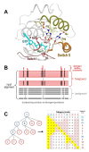
Figure 1
Figure 1
Structure and analysis of Arf/Arl GTPases. (A) Key structural features shared by Arf/Arl/Sar GTPases. In yellow are the side chains of six residues that are highly distinctive of Arf/Arl/Sar GTPases and that, within the GTP-bound state, often form the network of interactions shown. Within the Switch I region is shown a threonine residue (magenta side chain) that is conserved in the TRAFAC subclass of P-loop GTPases and that coordinates with the Mg++ ion associated with bound GTP. The structure shown is that of Arf1 bound to GTP (pdb_id: 1o3y; 1.50 Ǻ resolution) [57]. The Switch I and II regions (red and orange backbones, respectively) and the α3 helix (dark yellow backbone) are indicated. (B) Schematic representation of a contrast alignment, which reveals the sequence features distinguishing one group of proteins (termed the foreground) from related, evolutionarily-divergent proteins (termed the background). Red and gray horizontal bars represent sequences belonging to the foreground and background, respectively. Black vertical bars represent residues that are conserved in the foreground and white vertical bars represent corresponding non-conserved positions in the background. The histogram above these columns indicates the relative strengths of the selective constraints associated with evolutionary divergence of the foreground sequences from the background sequences. (C) Correspondence between a phylogenetic tree and a hyperpartition. A rooted tree is shown both as a graph and as a hyperpartition. In the hyperpartition, each column corresponds to one node in the tree such that the '+' rows in that column correspond to that node's subtree, which serves as the foreground, whereas the '-' rows in that column correspond to the rest of the parent node's subtree, which serves as the background; the remaining (non-participating) nodes in that column are labeled with an 'o'. Internal nodes (shown in blue) correspond to 'miscellaneous' subgroups, that is to sequences that are assigned to a subtree (e.g., a family), but not to a leaf node (e.g., a specific subfamily).
Figure 2
Figure 2
Hyperpartition for P-loop GTPases with an emphasis on Arf/Arl GTPases. The symbol '+', '-' or 'o' indicates that the subgroup is assigned to that category's foreground, background, or non-participating-sequence partition, respectively. Miscellaneous subgroups are italicized. Rejected sequences are those sampled into a random sequence set.
Figure 3
Figure 3
Contrast alignments showing distinguishing sequence features of Arf/Arl GTPases. This is the mcBPPS program output file corresponding to the Arf1-related subgroup (second row in Fig. 2); the sequences shown correspond to the seed alignment, which consists of Arf1 GTPases from distinct phyla. (A) Conserved residues distinguishing the TRAFAC subclass of P-loop GTPases from non-GTPases (column 3 in Fig. 2). (B) Conserved residues distinguishing Ras-like from other TRAFAC GTPases (column 6). (C) Conserved residues distinguishing Arf/Arl/Sar GTPases from other P-loop GTPases (except for Gα subunits)(column 12). (D) Conserved residues distinguishing typical Arf/Arl GTPases (i.e., excluding Arl6 and Arl8) from Sar and Gα GTPases (column 13). In each alignment, the Arf1 seed sequences are shown explicitly, whereas directly below these only the most conserved residue patterns in the foreground (relative to the background) are shown. Directly below this the corresponding weighted residue frequencies are shown (denoted by 'wt_res_freqs'). Weighted frequencies are given in integer tenths, where an '8', for example, indicates that the corresponding residue occurs in 80%-90% of the (foreground) sequences. Below this the conserved patterns and their weighted frequencies for the background sequences are shown (in gray). The selective constraints imposed on the foreground (relative to the background) at pattern positions are indicated by the histograms above each alignment.
Figure 4
Figure 4
Structural features associated with co-conserved residues that are distinctive of Arf/Arl/Sar GTPases and that form a network of salt bridges. Arf/Arl/Sar GTPases features correspond to column 12 in Fig. 2 and to the contrast alignment in Fig. 3C. See the legend to Fig. 1A for side chain and backbone color schemes. Shown in gray are the side chains of residues at an (unclassified) position within the α3-helix that weakly conserves an acidic amino acid which also participates in the Arf/Arl salt-bridge network. (A) Arf1 + GTP (pdb_id: 1o3y; 1.50 Ǻ)[57]. (B) Arf6 + GTP + Cholera toxin (not shown) (pdb_id: 2a5d; 1.80 Ǻ) [36]. (C) Arl1 + GTP + Golgin-245 Grip domain (not shown) (pdb_id: 1upt; 1.7 Ǻ) [37]. (D) Arl2 + GDP + PO43- and its effector PDE-δ (not shown) (pdb: 1ksh; 1.80 Ǻ) [38]. (E) Arl6 + GTP (pdb_id: 2 h57; 2.00 Ǻ) [Structural Genomics Consortium]. (F) Sar1 GTPase bound to a GTP analog (GppNHp)) and to the GAP protein Sec23 (pdb_id: 1m2o; 2.50 Ǻ) [41]; the side chain of the arginine-finger of Sec23 is shown in gray. (G) Gα bound to RGS4, GDP and AlF4 (transition state structure)(pdb_id: 1agr; 2.8 Ǻ) [58]. The side-chains of three residues that are specifically conserved in Gα subunits[6] (one of which is the arginine finger, Arg178) are shown in blue.
Figure 5
Figure 5
Structural locations of Arf/Arl co-conserved residues. (A-C) Conserved residues (green side-chains) distinguishing Arf/Arl from Sar and Gα GTPases (column 13 in Fig. 2; Fig. 3D). (A) The structure of Arf1 bound to GTP. The region shown corresponds to a β sheet adjacent to the Walker B region. Note that the asparagine of the N-[VI] motif (Asn52) forms a hydrogen bond with the Walker B aspartate (Asp67), whereas the valine (Val53) packs up against a tryptophan (Trp66) that directly precedes the aspartate. An Arf/Arl-conserved isoleucine (Ile74) packs against the backbone connecting the switch I threonine residue (Thr48) to the switch I glycine (Gly50; unlabeled in figure). (B) The structure of Arf1 bound to GDP. Note that the β-strand corresponding to the Walker B "W-D" motif has shifted to the left by two residue positions due to Arf's interswitch toggle and that the N-[VI] motif interactions are disrupted. (C) The structural location of the Arf/Arl conserved glutamine (Gln128) and alanine (Ala125) residues associated with the guanine binding loop (the NK-x-D motif). The glutamine may stabilize this loop by forming hydrogen bonds with backbone oxygen atoms on either side of this loop, as shown. (D-E). Structural locations of residues (blue side-chains) that are conserved in and distinctive of Arl8 GTPases (column 16 in Fig. 2; Fig. S4D in Additional File 1). Residue side chains corresponding to the N-[IV] motif (of Arf/Arl GTPases) are shown in green. Note that Arl8-specific conserved residues form alternative interactions in the two forms, suggesting that they play a role in the interswitch toggle. (D) A non-interswitch toggled form of Arl8-GDP (pdb_id: 1zd9; structural genomics consortium). (E) An interswitch toggled form of Arl8-GDP (pdb_id: 2 h18; structural genomics consortium).

Similar articles

Cited by

References

    1. Wittinghofer A. In: GTPases. Hall A, editor. Oxford: Oxford University Press; 2000. The Functioning of Molecular Switches in Three Dimensions; pp. 244–310.
    1. Walker JE, Saraste M, Runswick MJ, Gay NJ. Distantly related sequences in the alpha- and beta-subunits of ATP synthase, myosin, kinases and other ATP-requiring enzymes and a common nucleotide binding fold. Embo J. 1982;1:945–951. - PMC - PubMed
    1. Saraste M, Sibbald PR, Wittinghofer A. The P-loop--a common motif in ATP- and GTP-binding proteins. Trends Biochem Sci. 1990;15:430–434. doi: 10.1016/0968-0004(90)90281-F. - DOI - PubMed
    1. Leipe DD, Wolf YI, Koonin EV, Aravind L. Classification and evolution of P-loop GTPases and related ATPases. J Mol Biol. 2002;317:41–72. doi: 10.1006/jmbi.2001.5378. - DOI - PubMed
    1. Bhattacharyya RP, Remenyi A, Yeh BJ, Lim WA. Domains, motifs, and scaffolds: the role of modular interactions in the evolution and wiring of cell signaling circuits. Annu Rev Biochem. 2006;75:655–680. doi: 10.1146/annurev.biochem.75.103004.142710. - DOI - PubMed

Publication types

MeSH terms

LinkOut - more resources