. 2010 Dec 10;87(6):866-72.
doi: 10.1016/j.ajhg.2010.10.031.

PSMB8 encoding the β5i proteasome subunit is mutated in joint contractures, muscle atrophy, microcytic anemia, and panniculitis-induced lipodystrophy syndrome

Affiliations

PSMB8 encoding the β5i proteasome subunit is mutated in joint contractures, muscle atrophy, microcytic anemia, and panniculitis-induced lipodystrophy syndrome

Anil K Agarwal et al. Am J Hum Genet. .

Abstract

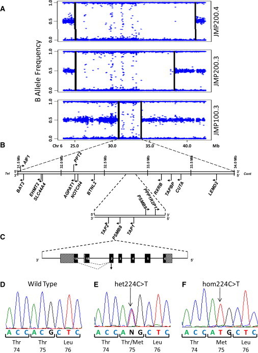
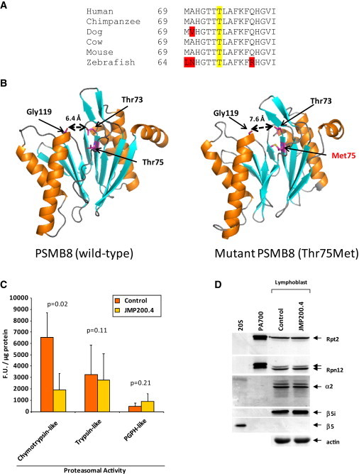
We performed homozygosity mapping in two recently reported pedigrees from Portugal and Mexico with an autosomal-recessive autoinflammatory syndrome characterized by joint contractures, muscle atrophy, microcytic anemia, and panniculitis-induced lipodystrophy (JMP). This revealed only one homozygous region spanning 2.4 Mb (5818 SNPs) on chromosome 6p21 shared by all three affected individuals from both families. We directly sequenced genes involved in immune response located in this critical region, excluding the HLA complex genes. We found a homozygous missense mutation c.224C>T (p.Thr75Met) in the proteasome subunit, beta-type, 8 (PSMB8) gene in affected patients from both pedigrees. The mutation segregated in an autosomal-recessive fashion and was not detected in 275 unrelated ethnically matched healthy subjects. PSMB8 encodes a catalytic subunit of the 20S immunoproteasomes called β5i. Immunoproteasome-mediated proteolysis generates immunogenic epitopes presented by major histocompatibility complex (MHC) class I molecules. Threonine at position 75 is highly conserved and its substitution with methionine disrupts the tertiary structure of PSMB8. As compared to normal lymphoblasts, those from an affected patient showed significantly reduced chymotrypsin-like proteolytic activity mediated by immunoproteasomes. We conclude that mutations in PSMB8 cause JMP syndrome, most probably by affecting MHC class I antigen processing.


Figures

Figure 1
Figure 1
Homozygous Region Shared by Affected Individuals with JMP Syndrome, Position of Candidate Genes in the Region, Structure of PSMB8, and Disease-Causing Mutation (A) Homozygous region shared by the affected individuals JMP 200.4, 200.3, and 100.3. The B allele frequency is a normalized measure of the allelic intensity ratio of the two alleles, and deviation from 0.5 indicates homozygosity. The genomic position is based on NCBI36/hg18 assembly. Each dot indicates the SNP genotyped by means of the Illumina HumanOmni1-Quad Beadchip. The vertical lines for each panel indicate the regions of homozygosity. Isolated heterozygous SNPs within larger homozygous segments are likely to represent miscalls. (B) Position of some of the candidate genes in the critical region identified by homozygosity mapping. Tel, telomeric end; cent, centromeric end. The critical region spanned from 31.5 Mb to 34.0 Mb on chromosome 6. (C) Structure of PSMB8 and the location of the mutation in the JMP patients. Human PSMB8 contains six exons and five introns. Also shown are the alternatively spliced exons 1A and 1B. The mutation c.224C>T in PSMB8 is located in exon 2. (D) Sequence electropherogram for normal sequence in exon 2 of PSMB8. (E) Sequence from JMP 200.2 showing heterozygous c.224C>T mutation. (F) Sequence from JMP 200.3 showing the homozygous c.224C>T mutation. The amino acid substitution is shown below the electropherogram.
Figure 2
Figure 2
Conservation of the Mutated Residue across Species, Crystal Structure of Normal and Mutant PSMB8, Proteasomal Activity, and Expression of β5i in Cell Lysates of Lymphoblasts from an Affected Patient (A) Alignment of partial PSMB8 amino acid sequence around threonine at position 75 in human (NP_683720.2), chimpanzee (XP_001167272.1), dog (XP_532100.1), cow (NP_001035570.1), mouse (NP_034854.1), and zebrafish (NP_571467.2). The conserved residue threonine is highlighted in yellow. Amino acids that are not conserved are shown in red. (B) Protein model for the wild-type and mutant, Thr75Met, human PSMB8. β sheets are shown in blue, helices in orange, and unstructured loops in gray. Threonine 73 and glycine 119 are the catalytic amino acids. Mutation of threonine 75 to methionine widens the distance between the catalytic threonine 73 and glycine 119 by 1.2 Å. All amino acids are numbered according to transcript 1B. (C) Proteasomal activity in EBV-transformed lymphoblasts from a control and an affected patient (JMP200.4). The chymotrypsin-like, trypsin-like, and PGPH-like proteolytic activities are expressed as fluorogenic units (F.U.) per μg of protein. Each bar represents mean ± SD from three independent experiments carried out in duplicate. p values were derived from the paired t test. (D) Western blots. Total cell lysates from EBV-transformed lymophoblasts of a control subject and an affected patient (JMP200.4) were resolved on 10% SDS-PAGE and probed with antibodies against Rpt2 (an ATPase of the PA700, a 19S proteasome regulatory particle), Rpn12 (a subunit of 19S proteasome regulatory subunit), α2, β5i, and β5 (subunits of 20S proteasome), and a housekeeping protein actin. The first two lanes on the left contain purified 20S and PA700 proteins from bovine red blood cells as positive controls. The control and the patient contained the same amount of β5i protein.

References

    1. Garg A., Hernandez M.D., Sousa A.B., Subramanyam L., de Villarreal L.M., Dos Santos H.G., Barboza O. An autosomal recessive syndrome of joint contractures, muscular atrophy, microcytic anemia, and panniculitis-associated lipodystrophy. J. Clin. Endocrinol. Metab. 2010;95:E58–E63. - PMC - PubMed
    1. Tanaka M., Miyatani N., Yamada S., Miyashita K., Toyoshima I., Sakuma K., Tanaka K., Yuasa T., Miyatake T., Tsubaki T. Hereditary lipo-muscular atrophy with joint contracture, skin eruptions and hyper-gamma-globulinemia: A new syndrome. Intern. Med. 1993;32:42–45. - PubMed
    1. Yamada S., Toyoshima I., Mori S., Tsubaki T. [Sibling cases with lipodystrophic skin change, muscular atrophy, recurrent skin eruptions, and deformities and contractures of the joints. A possible new clinical entity] Rinsho Shinkeigaku. 1984;24:703–710. - PubMed
    1. Oyanagi K., Sasaki K., Ohama E., Ikuta F., Kawakami A., Miyatani N., Miyatake T., Yamada S. An autopsy case of a syndrome with muscular atrophy, decreased subcutaneous fat, skin eruption and hyper gamma-globulinemia: Peculiar vascular changes and muscle fiber degeneration. Acta Neuropathol. 1987;73:313–319. - PubMed
    1. Horikoshi A., Iwabuchi S., Iizuka Y., Hagiwara T., Amaki I. [A case of partial lipodystrophy with erythema, dactylic deformities, calcification of the basal ganglia, immunological disorders, and low IQ level (author's transl)] Rinsho Shinkeigaku. 1980;20:173–180. - PubMed

Publication types

Substances