. 2010 Dec 10;87(6):768-78.
doi: 10.1016/j.ajhg.2010.11.005.

Loss of CHSY1, a secreted FRINGE enzyme, causes syndromic brachydactyly in humans via increased NOTCH signaling

Affiliations

Loss of CHSY1, a secreted FRINGE enzyme, causes syndromic brachydactyly in humans via increased NOTCH signaling

Jing Tian et al. Am J Hum Genet. .

Abstract

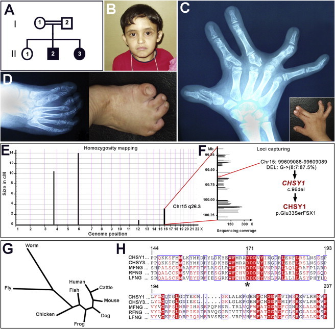
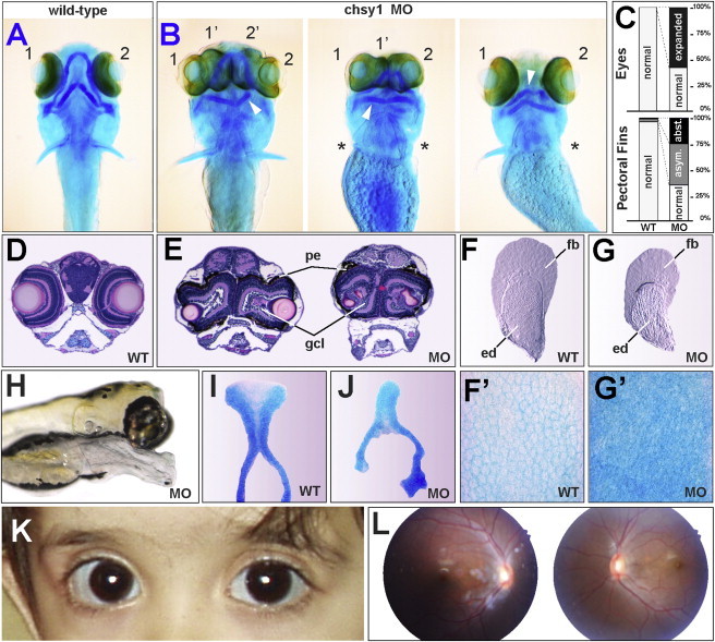
We delineated a syndromic recessive preaxial brachydactyly with partial duplication of proximal phalanges to 16.8 Mb over 4 chromosomes. High-throughput sequencing of all 177 candidate genes detected a truncating frameshift mutation in the gene CHSY1 encoding a chondroitin synthase with a Fringe domain. CHSY1 was secreted from patients' fibroblasts and was required for synthesis of chondroitin sulfate moieties. Noticeably, its absence triggered massive production of JAG1 and subsequent NOTCH activation, which could only be reversed with a wild-type but not a Fringe catalytically dead CHSY1 construct. In vitro, depletion of CHSY1 by RNAi knockdown resulted in enhanced osteogenesis in fetal osteoblasts and remarkable upregulation of JAG2 in glioblastoma cells. In vivo, chsy1 knockdown in zebrafish embryos partially phenocopied the human disorder; it increased NOTCH output and impaired skeletal, pectoral-fin, and retinal development. We conclude that CHSY1 is a secreted FRINGE enzyme required for adjustment of NOTCH signaling throughout human and fish embryogenesis and particularly during limb patterning.


Figures

Figure 1
Figure 1
Phenotypic Characteristics of Investigated Patients with Syndromic Recessive Brachydactyly Caused by a Frameshift Mutation in CHSY1 (A) Affected children, male (II:2) and female (II:3), were born to first-cousin parents (I:1 and I:2) with one unaffected child (II:1). (B) Head shot of affected boy (II:2) with protruding eyes, downward turn of mouth, and micrognathia. (C) X-ray radiograph of male proband's right hand when he was 8 years of age, showing partial duplications of proximal phalanges in digits 1, 2, and 3 (inset: hand photograph). (D) X-ray radiograph and picture of male proband's right foot showing severe skeletal anomalies. The big toe exhibits short and duplicated metatarsals and proximal phalanges. The second and fourth proximal phalanges are duplicated as well. (E) Homozygosity mapping delineated four candidate loci totaling 19 cM on chromosomes 3, 5, 12, and 15. (F) Genomic capturing and resequencing of candidate loci revealed a 1 bp deletion in CHSY1 on chromosome 15; this deletion caused an early-termination stop codon at amino acid 34 of the CHSY1 enzyme. (G) Phylogenetic tree showing conservation of CHSY1 across invertebrate and vertebrate species. (H) The CHSY1 enzyme and its paralogous protein CHSY3 bear a Fringe domain that aligns to that of canonical MANICAL (MFNG), RADICAL (RFNG), and LUNATIC (LFNG) FRINGE. A conserved DDD motif is highlighted with a star.
Figure 2
Figure 2
Loss of CHSY1 Results in Depletion of Chondroitin Sulfates and Activation of NOTCH Signaling (A) The full-length CHSY1 (WT) consists of a type II transmembrane domain, a Fringe motif, and a type A glycosyltransferase domain. The frameshift mutation (MUT) deletes more than 95% of CHSY1, including both of its putative catalytic sites, marked with an asterisk. (B) CHSY1 is found in the supernatant of primary fibroblast cultures from unaffected sibling II:1 (WT) but not from that of the affected proband II:2 (MUT). (C and D) Depletion of CS, a product of the CHSY1 enzyme, is observed by immunohistochemistry in the skin of affected II:2 (D) versus unaffected sibling II:1 (C). CHSY1-deficient primary fibroblasts (D, inset) have aberrant morphologies and slower growth rates than control fibroblasts (C, inset). (E) Massive upregulation of JAG1 and HES1 in affected primary fibroblasts, indicative of abnormally high NOTCH signaling, is observed by q-PCR analysis. Mutant CHSY1 transcripts were not subjected to nonsense-mediated decay. Upregulation of NOTCH signaling was reversed by the addition of a γ-secretase inhibitor. (F) Overexpression of intact CHSYI, but not of a Fringe-dead CHSY1 D171A mutant, can reverse NOTCH activation in CHSY1-deficient fibroblasts. (G) Abnormally high JAG1 protein production is seen in CHSY1-deficient fibroblasts (lanes 3 and 4) compared to control fibroblasts (lanes 1 and 2). Addition of a γ-secretase inhibitor rescues NOTCH activation measured by HES1 levels in CHSY1-deficient cells (compare lanes 3 and 4). Values in (E) and (F) represent means ± SE of data from three independent experiments, p < 0.05.
Figure 3
Figure 3
CHSY1 Knockdown Promotes NOTCH Signaling and Enhances Osteogenesis In Vitro (A) CHSY1 siRNA in human fetal osteoblasts (hFOB) cells achieved 75% knockdown with concurrent 6-fold upregulation of HES1, an immediate NOTCH target. Other genes belonging to the NOTCH pathway were moderately but significantly upregulated. (B) CHSY1 siRNA triggered upregulation of osteogenic markers, indicative of premature differentiation, whereas proliferation markers were downregulated. (C) CHSY1 siRNA in human glioblastoma cells (T98G) achieved 87% knockdown and a paralleled 10-fold upregulation of HES1 and 24-fold increase in JAG2 transcription. Values in (A)–(C) represent means ± SE of data from three independent experiments, p < 0.05.
Figure 4
Figure 4
chsy1 Knockdown in Zebrafish Embryos Affects Segmentation and Increases Notch Signaling (A) Six-day-old wild-type embryo. (B) Six-day-old chsy1 MO-injected embryo with a protruding mouth-piece, eyes joined at the midline, and arched trunk. (C) Six-day-old chsy1 MO-injected embryo rescued with 200 pg of human CHSY1 mRNA. (D) Ten-somite-stage wild-type embryo stained with pax2.1 marking the optic stalk (os), midbrain-hindbrain boundary (mhb), otic placodes (op), and pronephric duct (pd) and with papc marking the pre-somitic mesoderm (psm) and the tailbud (tb). (E) Ten-somite stage chsy1 morphant embryo displays a fused anterior eyefield, near-absent isthmic organizer, and otic placodes marked by pax2.1. Aberrant segmentation of the pre-somitic mesoderm and lack of papc staining in the tailbud indicate anomalies in somitogenesis. (F and F′) Wild-type (F) and chsy1 morphant (F′) 24 hpf embryos stained for the Notch ligand jag2. Note ectopic or delayed clearance of jag2 expression in trunk somites. (G and G′) Wild-type (G) and chsy1 morphant (G′) 24 hpf embryos stained for the Notch ligand deltaC. Note the enhanced expression in presomitic mesoderm at the tailbud level. (H and H′) Wild-type (H) and chsy1 morphant (H′) 24 hpf embryos that were stained with lim1 and whose expression is downregulated by Notch signaling. The loss of lim1 expression in head and trunk structures indicates greater-than-normal Notch signaling in chsy1 morphants relative to controls.
Figure 5
Figure 5
Phenotypes of chsy1 Zebrafish Morphant Embryos Partially Phenocopy Human Disorder (A) Ventral view of alcian-blue-stained 5-day-old wild-type embryo. (B) Three representative chsy1 MO-injected embryos exhibiting variable phenotypic penetrance, including skeletal anomalies (arrowheads), massive overgrowth of retinas, and pectoral-fin defects ranging from asymmetric development to unilateral or bilateral aplasia (asterisks). (C) Percentage of eyes and pectoral-fin defects scored in chsy1 MO compared to wild-type embryos. (Asym., asymmetric; Abst., absent). (D) Hematoxylin and Eosin (H&E) head sections of 5-day-old wild-type embryo. (E) H&E sections through midline-joining retinas of chsy1 morphant embryos at 5 days of development. Note hyperplasia of pigmented epithelium (pe) and increased cell numbers in the ganglion cell layer (gcl). (F and F′) Pectoral fin of a 6-day-old wild-type embryo. (F′) High-magnification view of the endoskeletal disc showing cartilage tissue with monolayered chondrocytes stained with alcian blue. (G and G′) Pectoral fin of a mildly affected 6-day-old chsy1 MO-injected embryo exhibiting hypoplasia of the endoskeletal disc (ed) relative to the fin blade (fb). (G′) High-magnification view of cartilage tissue with multilayered disorganized chondrocytes in the endoskeletal disc of chsy1 morphant embryo stained with alcian blue. (H) Lateral view of 5-day-old chsy1 morphant embryos with severe jaw hypoplasia and eye defects. (I) Ethmoid plate of a 5-day-old wild-type embryo stained with alcian blue. (J) Irregularly shaped ethmoid plate of a 5-day-old chsy1 morphant embryo. (K) Affected girl (II:3), showing bilateral macrophthalmia and blue sclera. (L) Color fundus photographs of affected girl (II:3) at 6 years of age. The girl had bilateral tilted eye discs and normal visual acuity in each eye.

References

    1. Temtamy S.A., Aglan M.S. Brachydactyly. Orphanet J. Rare Dis. 2008;3:15. - PMC - PubMed
    1. Lehmann K., Seemann P., Silan F., Goecke T.O., Irgang S., Kjaer K.W., Kjaergaard S., Mahoney M.J., Morlot S., Reissner C. A new subtype of brachydactyly type B caused by point mutations in the bone morphogenetic protein antagonist NOGGIN. Am. J. Hum. Genet. 2007;81:388–396. - PMC - PubMed
    1. Seemann P., Schwappacher R., Kjaer K.W., Krakow D., Lehmann K., Dawson K., Stricker S., Pohl J., Plöger F., Staub E. Activating and deactivating mutations in the receptor interaction site of GDF5 cause symphalangism or brachydactyly type A2. J. Clin. Invest. 2005;115:2373–2381. - PMC - PubMed
    1. Dathe K., Kjaer K.W., Brehm A., Meinecke P., Nürnberg P., Neto J.C., Brunoni D., Tommerup N., Ott C.E., Klopocki E. Duplications involving a conserved regulatory element downstream of BMP2 are associated with brachydactyly type A2. Am. J. Hum. Genet. 2009;84:483–492. - PMC - PubMed
    1. Lehmann K., Seemann P., Stricker S., Sammar M., Meyer B., Süring K., Majewski F., Tinschert S., Grzeschik K.H., Müller D. Mutations in bone morphogenetic protein receptor 1B cause brachydactyly type A2. Proc. Natl. Acad. Sci. USA. 2003;100:12277–12282. - PMC - PubMed

Publication types

Substances