Review
. 2011 Jan;34(1):41-50.
doi: 10.1016/j.tins.2010.10.002. Epub 2010 Dec 2.

Development, specification, and diversity of callosal projection neurons

Affiliations
Review

Development, specification, and diversity of callosal projection neurons

Ryann M Fame et al. Trends Neurosci. 2011 Jan.

Abstract

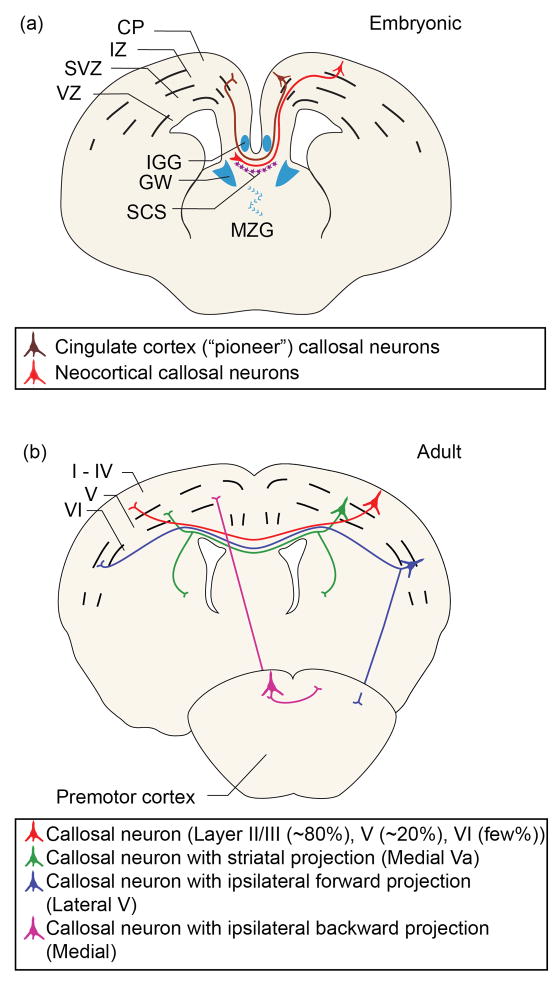
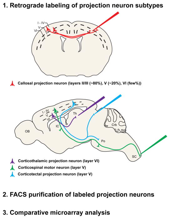
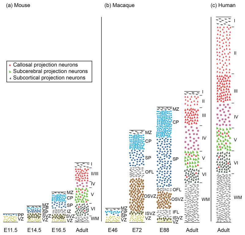
Callosal projection neurons (CPN) are a diverse population of neocortical projection neurons that connect the two hemispheres of the cerebral cortex via the corpus callosum. They play key roles in high-level associative connectivity, and have been implicated in cognitive syndromes of high-level associative dysfunction, such as autism spectrum disorders. CPN evolved relatively recently compared to other cortical neuron populations, and have undergone disproportionately large expansion from mouse to human. While much is known about the anatomical trajectory of developing CPN axons, and progress has been made in identifying cellular and molecular controls over midline crossing, only recently have molecular-genetic controls been identified that specify CPN populations, and help define CPN subpopulations. In this review, we discuss the development, diversity and evolution of CPN.


Figures

Figure 1
Figure 1. CPN development and diversity
(a) During development, callosal axons (red) turn toward the midline. Multiple glial populations (blue) and mixed neuronal/glial populations (purple) play critical roles in CPN axon guidance and midline crossing. Pioneering axons (brown) from neurons of the cingulate cortex begin the process of midline crossing. This schematic represents processes that occur across multiple embryonic times during mouse CPN development. Abbreviations: CP, cortical plate; IZ, intermediate zone; SVZ, subventricular zone; VZ, ventricular zone; IGG, indusium griseum glia; GW, glial wedge; SCS, subcallosal sling; MZG, midline zipper glia. (b) At least four major types of adult CPN can be classified based on projection patterns. These include: single projections to the contralateral cortex (red); dual projections to the contralateral cortex and ipsilateral or contralateral striatum (green); dual projections to the contralateral cortex and ipsilateral premotor cortex (blue); or dual projections to the contralateral cortex and ipsilateral sensorimotor cortex (purple).
Figure 2
Figure 2
Schematic representation of an experimental approach used to identify CPN-specific genes. CPN (red), corticothalamic projection neurons (purple), corticospinal motor neurons (green), and corticotectal projection neurons (blue) were retrogradely labeled at distinct stages of development from the contralateral hemisphere, the thalamus, the spinal cord, and the superior colliculus, respectively. Labeled neurons were dissociated, purified using fluorescence activated cell sorting (FACS), and followed by comparative microarray genetic expression analysis[, , –84]. Adapted, with permission, from Ref. [81].
Figure 3
Figure 3. Spatially-restricted genes identify novel CPN subpopulations
Schematic representation of neocortical layers depicting laminar-specific expression of 20 selected, representative genes expressed by early postnatal CPN within the neocortex [81]. Dark and light blue bands indicate high and low levels of expression, respectively. Grey oblique stripes demarcate layers in which CPN reside. Most of these genes have dynamic patterns of expression through development; therefore, developmental stage must be considered when using these genes to identify specific populations of CPN. Representative genes are depicted with multiple patterns of laminar expression: most cortical layers; and deep cortical layers only (top row); superficial cortical layers only (middle row); and subdivisions of superficial layers (bottom row). See text and Table S1 in supplementary material online for more detailed expression and references. Abbreviations: roman numerals indicate neocortical layers (I-VI); SP, subplate.
Figure 4
Figure 4. Comparison of developing and adult mammalian neocortex of mouse, macaque, and human shows correlations between SVZ expansion, superficial neocortical layer expansion, and white matter expansion
Schematic comparison of histological sections of (a) developing and adult mouse, (b) developing and adult rhesus macaque monkey, and (c) adult human neocortex. Adult cross-sections are from visual cortex. The thicknesses are represented relative to a common scale. Callosal projection neurons (red) reside mostly in layers II/III (~80%), V (~20%), and VI (few %) in the adult neocotex. There is a strong correlation between the expansion of the SVZ and the expansion of the superficial layer thickness and neocortical white matter. Abbreviations: E, embryonic; PP, preplate; VZ, ventricular zone; MZ, marginal zone; CP, cortical plate; SP, subplate; SVZ, subventricular zone; OSVZ, outer SVZ; ISVZ, inner SVZ; OFL, outer fibrous layer; IFL, inner fibrous layer; WM, white matter; Roman numerals denote neocortical layers (IVI). Adapted and expanded with permission from Ref. [91] (macaque) with data from Ref [100] (mouse), and Refs [1, 99] (human).

References

    1. Schoenemann PT, et al. Prefrontal white matter volume is disproportionately larger in humans than in other primates. Nat Neurosci. 2005;8:242–252. - PubMed
    1. Aboitiz F, Montiel J. One hundred million years of interhemispheric communication: the history of the corpus callosum. Braz J Med Biol Res. 2003;36:409–420. - PubMed
    1. Peters A, Jones EG, editors. Cellular Components of the Cerebral Cortex. Plenum Press; 1984.
    1. Kowalczyk T, et al. Intermediate neuronal progenitors (basal progenitors) produce pyramidal-projection neurons for all layers of cerebral cortex. Cereb Cortex. 2009;19:2439–2450. - PMC - PubMed
    1. Noctor SC, et al. Cortical neurons arise in symmetric and asymmetric division zones and migrate through specific phases. Nat Neurosci. 2004;7:136–144. - PubMed

Publication types