. 2011 Feb 1;489(1):43-7.
doi: 10.1016/j.neulet.2010.11.063. Epub 2010 Dec 2.

Essential role of toll-like receptor 2 in morphine-induced microglia activation in mice

Affiliations

Essential role of toll-like receptor 2 in morphine-induced microglia activation in mice

Yi Zhang et al. Neurosci Lett. .

Abstract

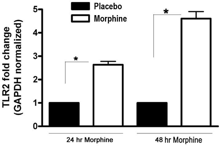
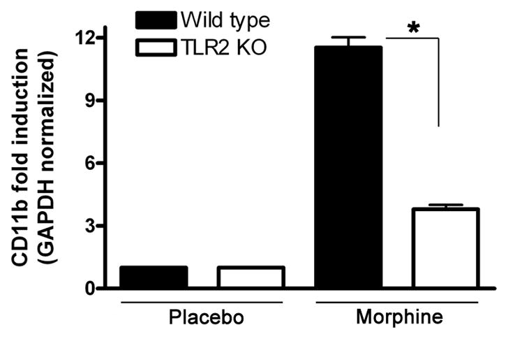
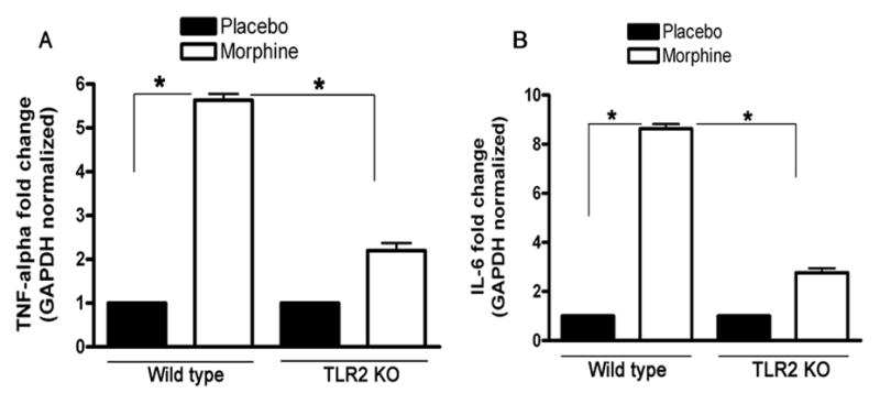
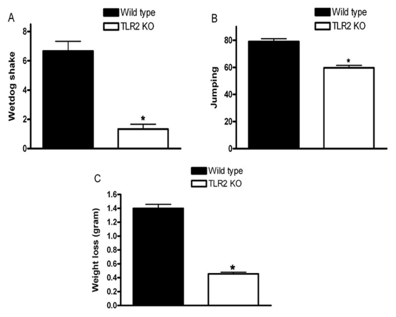
Opioids are powerful pain relievers, but also potent inducers of dependence and tolerance. Chronic morphine administration (via subcutaneous pellet) induces morphine dependence in the nucleus accumbens, an important dependence region in the brain, yet the cellular mechanisms are mostly unknown. Toll-like receptor 2 (TLR2) plays an essential function in controlling innate and inflammatory responses. Using a knockout mouse lacking TLR2, we assessed the contribution of TLR2 to microglia activation and development of morphine dependence. We report here that mice deficient in TLR2 inhibit morphine-induced the levels of microglia activation and proinflammatory cytokines. Moreover, in TLR2 knockout mice the main symptoms of morphine withdrawal were significantly attenuated. Our data reveal that TLR2 plays a critical role in morphine-induced microglia activation and dependence.


Figures

Fig. 1
Fig. 1
Chronic morphine administration induces the expression of TLR2. Wild type C57BL/6 male mice aged 9–10 week were implanted with a 25 mg sustained-release morphine pellet in the dorsal subcutaneous space, while control mice received a comparable placebo pellet as described under materials and methods. At 24 and 48 h after implantation, the mice were euthanized and the NAc was harvested and the expression of TLR2 was determined by real time quantitative RT-PCR. There were five mice per group. Means and SEs were calculated from 5 mice per group. * p < 0.01 compared with wild type placebo group.
Fig. 2
Fig. 2
TLR2 is required for the activation of microglia following chronic morphine administration. Wild type (WT) and TLR2 KO male mice aged 9–10 week were implanted with a 25 mg morphine pellet or a placebo pellet subcutaneously under isoflurane anesthesia as described under materials and methods. Four days after pellet implantation, the NAcs were harvested and total RNA was isolated. The activation level of microglia was quantified by measuring the transcript levels of CD11b by real time RT-PCR. Means and SEs were calculated from 5 to 7 mice per group. * p < 0.01 compared with indicated groups.
Fig. 3
Fig. 3
Effect of chronic administration of morphine on the expression of TNF-α, IFN-γ and IL-6 in wild type mice and TLR2 KO mice. 9–10-week-old TLR2 KO male mice (n = 5) and age-matched wild type male mice (n = 6) were implanted with a 25 mg morphine pellet or a placebo pellet subcutaneously under isoflurane anesthesia as described under materials and methods. At 72 h after pellet implantation, the NAcs were harvested and total RNA was isolated. The expression of TNF-α (A) and IL-6 (B) was determined by real time quantitative RT-PCR. The level of gene expression is presented as relative mRNA expression units. Means and SEs were calculated from 5 mice per group. ** p < 0.01 compared with indicated groups.
Fig. 4
Fig. 4
Naloxone-precipitated withdrawal after 72 h of morphine pellet implantation. 9–10-week-old TLR2 KO male mice and age-matched wild type male mice were implanted with a 25 mg morphine pellet subcutaneously or received a placebo pellet under isoflurane anesthesia as described under materials and methods. At 72 h after pellet implantation, mice received naloxone (1.0 mg/kg, i.p.) and morphine withdrawal symptoms for wet dog shakes (A), jumps (B), and weight loss (C) were monitored for 30 min. The weight of each mouse was determined before and 30 min after the naloxone injection. Means and SEs were calculated from 6 to 8 mice per group. * p < 0.01 compared with WT mice.

Similar articles

Cited by

References

    1. Yin D, Mufson RA, Wang R, Shi Y. Fas-mediated cell death promoted by opioids. Nature. 1999;397:218. - PubMed
    1. Yin D, Woodruff M, Zhang Y, Whaley S, Miao J, Ferslew K, Zhao J, Stuart C. Morphine promotes Jurkat cell apoptosis through pro-apoptotic FADD/P53 and anti-apoptotic PI3K/Akt/NF-kappaB pathways. J Neuroimmunol. 2006;174:101–107. - PubMed
    1. Moorman J, Zhang Y, Liu B, LeSage G, Chen Y, Stuart C, Prayther D, Yin D. HIV-1 gp120 primes lymphocytes for opioid-induced, beta-arrestin 2-dependent apoptosis. Biochim Biophys Acta. 2009;1793:1366–1371. - PMC - PubMed
    1. Holan V, Zajicova A, Krulova M, Blahoutova V, Wilczek H. Augmented production of proinflammatory cytokines and accelerated allotransplantation reactions in heroin-treated mice. Clin Exp Immunol. 2003;132:40–45. - PMC - PubMed
    1. Avila AH, Alonzo NC, Bayer BM. Immune cell activity during the initial stages of withdrawal from chronic exposure to cocaine or morphine. J Neuroimmunol. 2004;147:109–113. - PubMed

Publication types

LinkOut - more resources