Review
. 2011 Apr;23(2):126-34.
doi: 10.1016/j.ceb.2010.11.001. Epub 2010 Dec 3.

Modeling general proteostasis: proteome balance in health and disease

Affiliations
Review

Modeling general proteostasis: proteome balance in health and disease

Daniela M Roth et al. Curr Opin Cell Biol. 2011 Apr.

Abstract

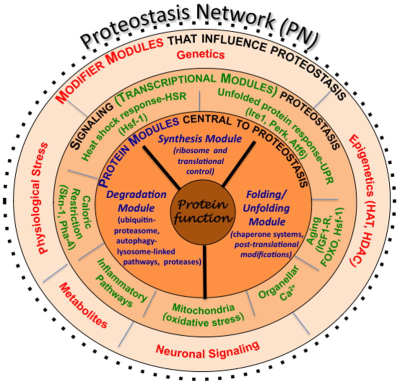
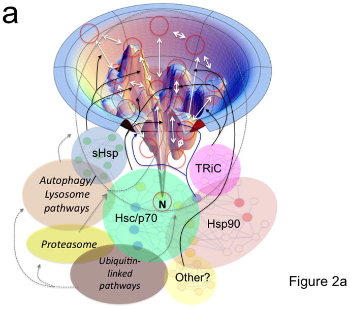
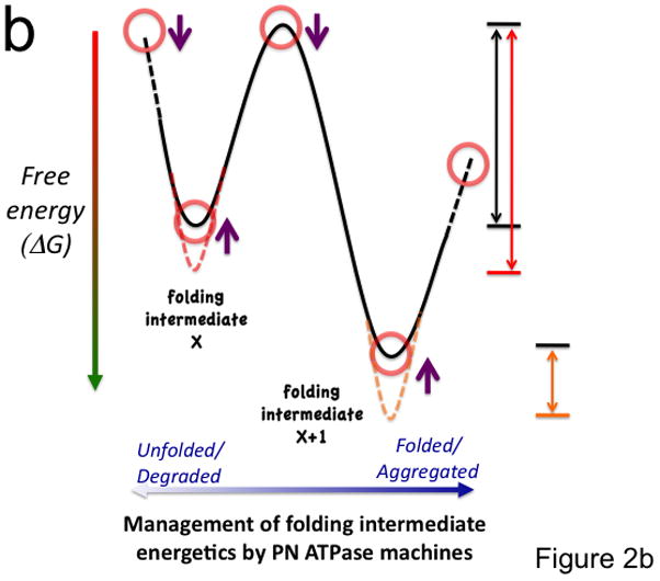
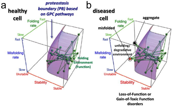
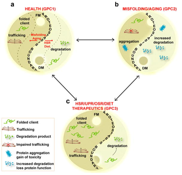
Protein function is generated and maintained by the proteostasis network (PN) (Balch et al. (2008) Science, 319:916). The PN is a modular, yet integrated system unique to each cell type that is sensitive to signaling pathways that direct development and aging, and respond to folding stress. Mismanagement of protein folding and function triggered by genetic, epigenetic and environmental causes poses a major challenge to human health and lifespan. Herein, we address the impact of proteostasis defined by the FoldFx model on our understanding of protein folding and function in biology. FoldFx describes how general proteostasis control (GPC) enables the polypeptide chain sequence to achieve functional balance in the context of the cellular proteome. By linking together the chemical and energetic properties of the protein fold with the composition of the PN we discuss the principle of the proteostasis boundary (PB) as a key component of GPC. The curved surface of the PB observed in 3-dimensional space suggests that the polypeptide chain sequence and the PN operate as an evolutionarily conserved functional unit to generate and sustain protein dynamics required for biology. Modeling general proteostasis provides a rational basis for tackling some of the most challenging diseases facing mankind in the 21st century.


Figures

Figure 1
Figure 1. The PN
Shown are the interactions that comprise the PN, the composition of which is responsible for generating and maintaining the biological protein fold. Components comprising the PN outlined in the inner-most layer (in blue font) involve the synthesis module, the folding/unfolding module, and the degradation module (the GPC triad). A second layer shows the signaling transcriptional pathways (in green font) that influence the level and activity of the triad found in the innermost layer. The third layer (in red font) includes modifiers that influence and/or integrate the activities defined by the second and first layers. Modifiers and signaling pathways from both cell autonomous and cell non-autonomous origin. Modified figure reproduced with permission from Elsevier Press [2].
Figure 2
Figure 2. Coupling of the folding energy landscape with the PN
(a) Illustrated is a bumpy energy landscape funnel (http://www.dillgroup.ucsf.edu/) in which an unfolded protein proceeds along various intermediate steps (red circles) that can pose energetic barriers to achieve the native state (N) at the base of the funnel. The white arrows indicate potential pathways through various folding intermediates that the nascent protein may take to reach the native state. The solid black and blue (synthesis and folding modules) and the dashed gray (degradative module) arrows illustrate how different components of the PN may influence pathway choice. The dashed oval indicates that all intermediates are potential steps in which a protein can be targeted for degradation. Hsp90 is thought to principally facilitate late folding events (solid blue lines). (b) The energy landscape (a slice through the funnel illustrated in panel a) illustrates the central role of the ATPase cycle in synthesis, folding and degradative modules in managing the biology of the protein structure in the cell to achieve function. By coupling the energy of ATP hydrolysis (X-axis) by PN ATPase machines with the energetics imparted in the chemistry of the amino acid sequence of the polypeptide chain (Y-axis), PN ATPases maintain the protein fold in a dynamic state- essential for biology. Right side of panel illustrates energy barriers necessary to achieve a functional fold (black arrow) relative to the energetics associated with misfolding (red and orange arrows). The additional energetic demands challenging GPC through misfolding (red line) or aggregation (short orange line) are illustrated. Purple arrows indicate potential steps for GPC to alter folding kinetics and energetics to promote proteome balance and cell health. Abbreviations: sHsp (small heat shock proteins); TRiC (TCP1-ring complex).
Figure 3
Figure 3. The proteostasis boundary (PB)
The position of each node (protein) relative to the PB (curved surface) responsible for biological function is defined by a protein’s folding properties: folding kinetics (Z-axis), misfolding kinetics (Y-axis) and thermodynamic stability (X-axis). Each line defines a physical or functional interaction between two proteins in the system. The location of the PB in 3D space is established by the composition of the PN and modulated by the GPC triad. (a) All of the nodes are within the PB boundary in a healthy cell. (b) Mutations or aberrant post-translational modifications can alter folding kinetics and energetics, making their corresponding nodes and edges fall outside (above the curved surface) of the PB. This space in the 3D plot does not support function of the energetically destabilized variant and can lead to either degradation (red node) or protein aggregation (black node). The loss of connectivity to proteins within the embrace of the PB can challenge the entire PN leading to cell, tissue, and organismal disease. Reproduced with permission from Elsevier Press [2].
Figure 4
Figure 4. Modeling the Yin-Yang of proteome balance in health, disease, and in response to proteostasis therapeutics
(Panel a) Proteome balance [4] in a healthy cell is determined by the composition of the synthesis and folding modules (FM) (the Yang on the left side of diagram) and degradative module (DM) (the Yin on the right side of diagram). GPC1 determines the position of the PB (the S-shaped curve) and healthspan. The dashed lines illustrate that misfolding and aging can challenge the position of the PB. (Panel b) Aging and unfolding move the PB to the left resulting in compromised proteostasis function (GPC2) and an unhealthy cell by triggering increased degradation and/or accumulation of protein aggregates. (Panel c) Biological signaling pathways including the HSR (HSF1 and IGF1-R/FOXO pathways), UPR, oxidative stress response (OSR), diet, IGF1-R and/or proteostasis targeted therapeutics can move the Yin-Yang balance defined by the PB to the right generating GPC3. GPC3 provides an environment that protects the cell from physiological stress, misfolding and aging, allowing the cell to return to GPC1.

Similar articles

Cited by

References

    1. Balch WE, Morimoto RI, Dillin A, Kelly JW. Adapting proteostasis for disease intervention. Science. 2008;319:916–919. - PubMed
    1. Powers ET, Morimoto RI, Dillin A, Kelly JW, Balch WE. Biological and chemical approaches to diseases of proteostasis deficiency. Annu Rev Biochem. 2009;78:959–991. This article develops in detail the concept of the FoldFx model for understanding the impact of proteostasis on human biology. - PubMed
    1. Douglas PM, Dillin A. Protein homeostasis and aging in neurodegeneration. J Cell Biol. 2010;190:719–729. - PMC - PubMed
    1. Hutt D, Balch WE. Cell Biology. The proteome in balance. Science. 2010;329:766–767. A short perspective on the article by Okiyoneda et al. (2010) (reference 32) that establishes the importance of proteostasis pathways affecting both synthesis and maintenance of the protein fold for function leading to proteome balance. - PMC - PubMed
    1. Morimoto RI. Proteotoxic stress and inducible chaperone networks in neurodegenerative disease and aging. Genes Dev. 2008;22:1427–1438. - PMC - PubMed