

Sensitivity of cytopathological examination in the diagnosis of feline sporotrichosis
- PMID: 21131220
- PMCID: PMC10832817
- DOI: 10.1016/j.jfms.2010.10.007
Sensitivity of cytopathological examination in the diagnosis of feline sporotrichosis
Abstract
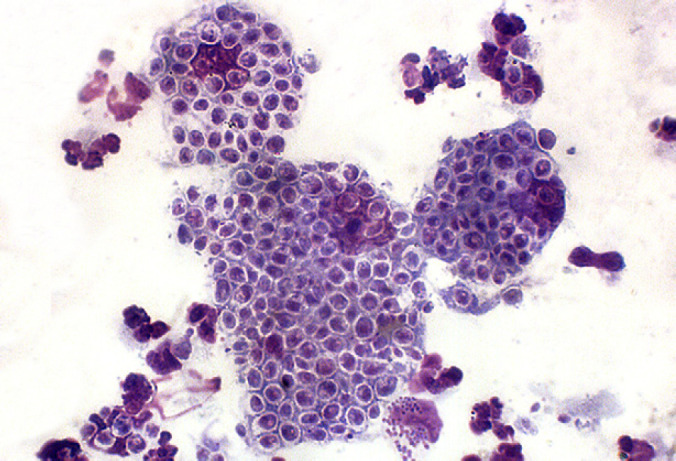
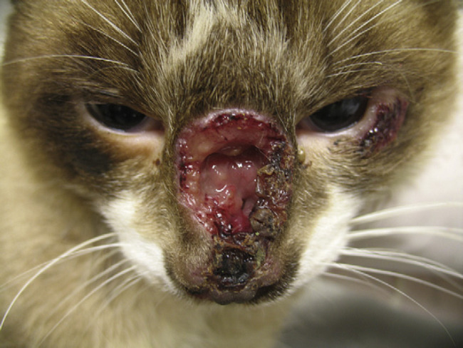
Sporotrichosis is caused by Sporothrix schenckii. The cat is the animal species most affected by this mycosis and plays an important role in the zoonotic transmission of this disease. The definitive diagnosis is made by isolation of the fungus in culture; however, cytopathological examination is frequently used in cats. Medical records from cats treated at Instituto de Pesquisa Clínica Evandro Chagas/Fiocruz, Rio de Janeiro, Brazil, between 2004 and 2006 were reviewed. Criteria for inclusion were a diagnosis by isolation of S schenckii in culture and cytopathological examination of the same lesion as that used for collection of the culture material. Eight hundred and six cats were included in the study. Yeast-like structures suggestive of S schenckii were observed in 636 cases. The sensitivity of the method was 78.9%. Its practicality, low cost and sensitivity validate cytopathology as a safe technique for the presumptive diagnosis of sporotrichosis in cats.
Copyright © 2010 ISFM and AAFP. Published by Elsevier Ltd. All rights reserved.
Figures
References
-
- Kauffman C.A. Sporotrichosis, Clin Infect Dis 29, 1999, 231–236. - PubMed
-
- Schubach A., Barros M.B., Wanke B. Epidemic sporotrichosis, Current Opin Infect Dis 21, 2008, 129–133. - PubMed
-
- Pereira S.A., Passos S.R.L., Silva J.N., et al. Response to azolic antifungal agents for treating feline sporotrichosis, Vet Rec 166, 2010, 290–294. - PubMed
-
- Ramos-e-Silva M., Vasconcelos C., Carneiro S., Cestari T. Sporotrichosis, Clin Dermatol 25, 2007, 181–187. - PubMed
-
- Costa E.O., Diniz L.S., Netto C.F., Arruda C., Dagli M.L. Epidemiological study of sporotrichosis and histoplasmosis in captive Latin American wild mammals, Sao Paulo, Brazil, Mycopathologia 125, 1994, 19–22. - PubMed
Publication types
MeSH terms
LinkOut - more resources
Full Text Sources
Medical
Miscellaneous
