

Generalized 3-Hz spike-and-wave complexes emanating from focal epileptic activity in pediatric patients
- PMID: 21131239
- PMCID: PMC3992252
- DOI: 10.1016/j.yebeh.2010.10.025
Generalized 3-Hz spike-and-wave complexes emanating from focal epileptic activity in pediatric patients
Abstract
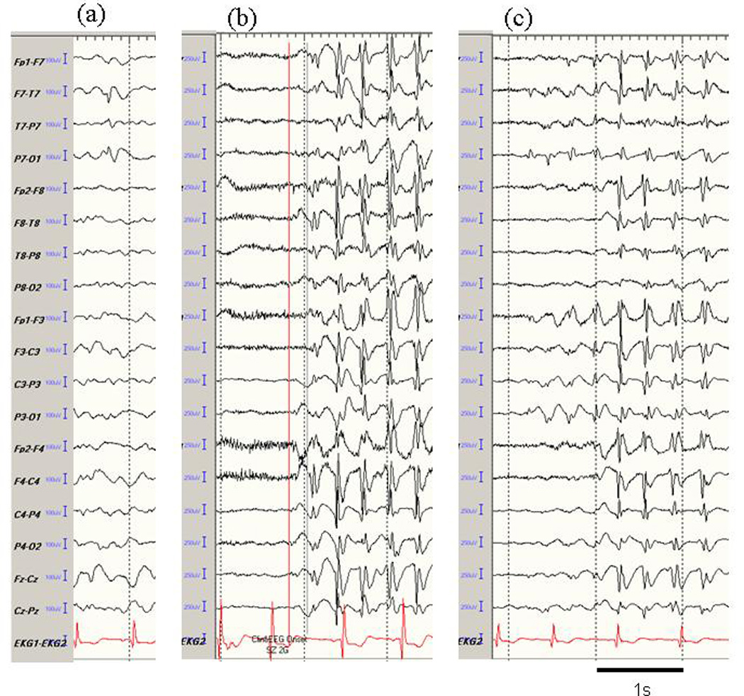
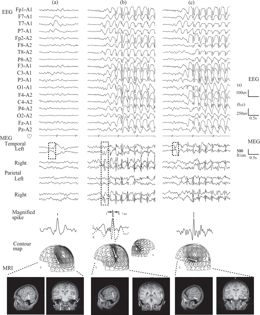
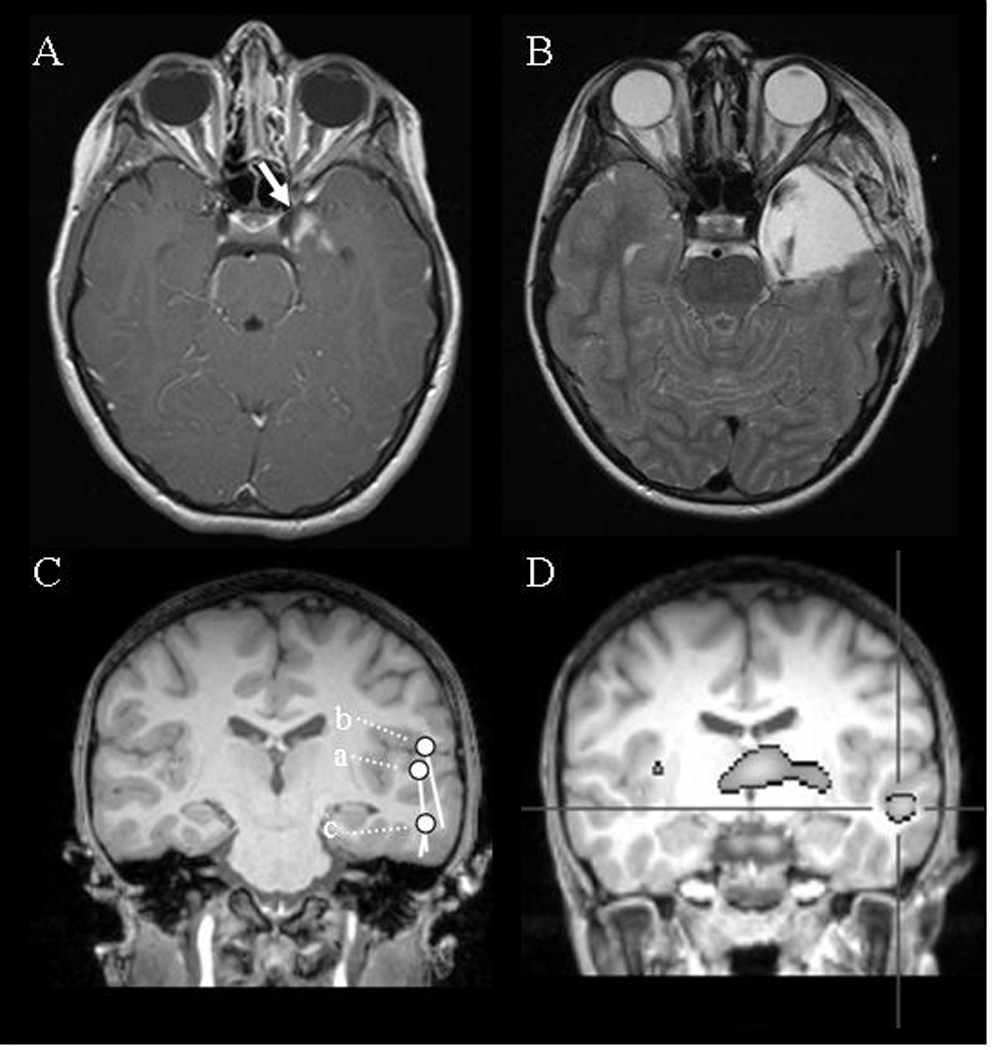
We describe two pediatric patients with an uncommon electrophysiological seizure propagation pattern. Both had dialeptic seizures as the main or only symptom. Case 1 had a small mass in the left medial temporal structures; case 2 had no lesion on magnetic resonance imaging. In both, the electroencephalogram showed not only left temporal spikes, but also bilaterally synchronous 3-Hz spike-and-wave complexes (SWCs) from onset and unusual secondarily generalized 3-Hz SWC patterns arising from the left temporal region. Case 1 was seizure free following resection of the mass; focal or generalized epileptiform electroencephalographic abnormalities were no longer present. In case 2, magnetoencephalography localized the spikes to the left superior and midtemporal gyrus, which ictal single-photon-emission computed tomography suggested was the origin of onset. These cases illustrate the close relationship between the focal epileptic area and 3-Hz SWCs and suggest that the focal area can trigger 3-Hz SWCs. The therapeutic strategy may need to be altered in such patients.
Copyright © 2010 Elsevier Inc. All rights reserved.
Figures
Similar articles
-
Magnetoencephalographic Identification of Epileptic Focus in Children With Generalized Electroencephalographic (EEG) Features but Focal Imaging Abnormalities.J Child Neurol. 2017 Oct;32(12):981-995. doi: 10.1177/0883073817724903. Epub 2017 Aug 22. J Child Neurol. 2017. PMID: 28828916
-
Magnetoencephalography localizing spike sources of atypical benign partial epilepsy.Brain Dev. 2014 Jan;36(1):21-7. doi: 10.1016/j.braindev.2012.12.011. Epub 2013 Feb 4. Brain Dev. 2014. PMID: 23384398
-
Ictal single photon emission computed tomography in absence seizures: apparent implication of different neuronal mechanisms.J Child Neurol. 2001 May;16(5):339-44. doi: 10.1177/088307380101600506. J Child Neurol. 2001. PMID: 11392518
-
Epileptiform electroencephalographic patterns.Mayo Clin Proc. 1996 May;71(5):501-11. doi: 10.4065/71.5.501. Mayo Clin Proc. 1996. PMID: 8628033 Review.
-
Is there such a thing as "generalized" epilepsy?Adv Exp Med Biol. 2014;813:81-91. doi: 10.1007/978-94-017-8914-1_7. Adv Exp Med Biol. 2014. PMID: 25012369 Review.
Cited by
-
Contributions of Magnetoencephalography to Understanding Mechanisms of Generalized Epilepsies: Blurring the Boundary Between Focal and Generalized Epilepsies?Front Neurol. 2022 Apr 27;13:831546. doi: 10.3389/fneur.2022.831546. eCollection 2022. Front Neurol. 2022. PMID: 35572923 Free PMC article. Review.
-
Genetic generalized epilepsies with frontal lesions mimicking migratory disorders on the epilepsy monitoring unit.Epilepsia Open. 2020 Mar 12;5(2):176-189. doi: 10.1002/epi4.12385. eCollection 2020 Jun. Epilepsia Open. 2020. PMID: 32524043 Free PMC article.
-
Magnetoencephalography in pediatric epilepsy.Korean J Pediatr. 2013 Oct;56(10):431-8. doi: 10.3345/kjp.2013.56.10.431. Epub 2013 Oct 31. Korean J Pediatr. 2013. PMID: 24244211 Free PMC article. Review.
-
Focal Epileptogenic Lesions in Adult Patients with Epilepsy and Generalized Epileptiform Discharges.J Epilepsy Res. 2016 Dec 31;6(2):75-78. doi: 10.14581/jer.16014. eCollection 2016 Dec. J Epilepsy Res. 2016. PMID: 28101478 Free PMC article.
-
Seizure Outcome and Its Prognostic Predictors After Hemispherotomy in Children With Refractory Epilepsy in a Chinese Pediatric Epileptic Center.Front Neurol. 2019 Aug 14;10:880. doi: 10.3389/fneur.2019.00880. eCollection 2019. Front Neurol. 2019. PMID: 31474931 Free PMC article.
References
-
- Commission on Classification and Terminology of the International League Against Epilepsy. Proposal for revised classification of epilepsies and epileptic syndromes. Epilepsia. 1989;30:389–399. - PubMed
-
- Meeren H, van Luijtelaar G, Lopes da Silva F, Coenen A. Evolving concepts on the pathophysiology of absence seizures: the cortical focus theory. Arch Neurol. 2005;62:371–376. - PubMed
-
- Gupta A, Chirla A, Wyllie E, Lachhwani DK, Kotagal P, Bingaman WE. Pediatric epilepsy surgery in focal lesions and generalized electroencephalogram abnormalities. Pediatr Neurol. 2007;37:8–15. - PubMed
-
- Luders H, Lesser RP, Dinner DS, Morris HH., 3rd Generalized epilepsies: a review. Cleve Clin Q. 1984;51:205–226. - PubMed
-
- Wyllie E, Lachhwani DK, Gupta A, Chirla A, Cosmo G, Worley S, et al. Successful surgery for epilepsy due to early brain lesions despite generalized EEG findings. Neurology. 2007;69:389–397. - PubMed
