

Discovery of very late antigen-4 (VLA-4, alpha4beta1 integrin) allosteric antagonists
- PMID: 21131351
- PMCID: PMC3037658
- DOI: 10.1074/jbc.M110.162636
Discovery of very late antigen-4 (VLA-4, alpha4beta1 integrin) allosteric antagonists
Abstract
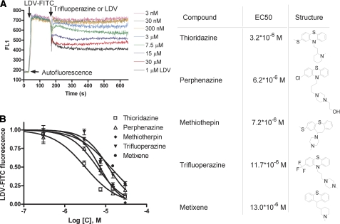
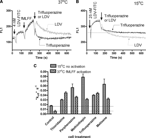
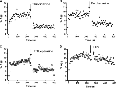
Integrins are cell adhesion receptors that mediate cell-to-cell, or cell-to-extracellular matrix adhesion. They represent an attractive target for treatment of multiple diseases. Two classes of small molecule integrin inhibitors have been developed. Competitive antagonists bind directly to the integrin ligand binding pocket and thus disrupt the ligand-receptor interaction. Allosteric antagonists have been developed primarily for α(L)β(2)- integrin (LFA-1, lymphocyte function-associated antigen-1). Here we present the results of screening the Prestwick Chemical Library using a recently developed assay for the detection of α(4)β(1)-integrin allosteric antagonists. Secondary assays confirmed that the compounds identified: 1) do not behave like competitive (direct) antagonists; 2) decrease ligand binding affinity for VLA-4 ∼2 orders of magnitude; 3) exhibit antagonistic properties at low temperature. In a cell based adhesion assay in vitro, the compounds rapidly disrupted cellular aggregates. In accord with reports that VLA-4 antagonists in vivo induce mobilization of hematopoietic progenitors into the peripheral blood, we found that administration of one of the compounds significantly increased the number of colony-forming units in mice. This effect was comparable to AMD3100, a well known progenitor mobilizing agent. Because all the identified compounds are structurally related, previously used, or currently marketed drugs, this result opens a range of therapeutic possibilities for VLA-4-related pathologies.
Figures
Similar articles
-
Small molecule agonist of very late antigen-4 (VLA-4) integrin induces progenitor cell adhesion.J Biol Chem. 2013 Jul 5;288(27):19414-28. doi: 10.1074/jbc.M113.479634. Epub 2013 May 23. J Biol Chem. 2013. PMID: 23703610 Free PMC article.
-
Decreased stroma adhesion capacity of CD34+ progenitor cells from mobilized peripheral blood is not lineage- or stage-specific and is associated with low beta 1 and beta 2 integrin expression.J Hematother Stem Cell Res. 2002 Jun;11(3):491-500. doi: 10.1089/15258160260090951. J Hematother Stem Cell Res. 2002. PMID: 12183834
-
Galphas-coupled receptor signaling actively down-regulates alpha4beta1-integrin affinity: a possible mechanism for cell de-adhesion.BMC Immunol. 2008 Jun 5;9:26. doi: 10.1186/1471-2172-9-26. BMC Immunol. 2008. PMID: 18534032 Free PMC article.
-
VLA-4-mediated interactions between normal human hematopoietic progenitors and stromal cells.Leuk Lymphoma. 1997 Feb;24(5-6):423-35. doi: 10.3109/10428199709055581. Leuk Lymphoma. 1997. PMID: 9086434 Review.
-
Mobilization of hematopoietic stem and progenitor cells using inhibitors of CXCR4 and VLA-4.Leukemia. 2012 Jan;26(1):34-53. doi: 10.1038/leu.2011.197. Epub 2011 Sep 2. Leukemia. 2012. PMID: 21886173 Free PMC article. Review.
Cited by
-
A Flow Cytometry-Based High-Throughput Technique for Screening Integrin-Inhibitory Drugs.J Vis Exp. 2024 Feb 2;(204):10.3791/64401. doi: 10.3791/64401. J Vis Exp. 2024. PMID: 38372326 Free PMC article.
-
Integrin-based therapeutics: biological basis, clinical use and new drugs.Nat Rev Drug Discov. 2016 Mar;15(3):173-83. doi: 10.1038/nrd.2015.10. Epub 2016 Jan 29. Nat Rev Drug Discov. 2016. PMID: 26822833 Free PMC article. Review.
-
Drug Repurposing from an Academic Perspective.Drug Discov Today Ther Strateg. 2011 Winter;8(3-4):61-69. doi: 10.1016/j.ddstr.2011.10.002. Drug Discov Today Ther Strateg. 2011. PMID: 22368688 Free PMC article.
-
In vitro and in vivo effects of AVA4746, a novel competitive antagonist of the ligand binding of VLA-4, in B-cell acute lymphoblastic leukemia.Exp Ther Med. 2022 Jan;23(1):47. doi: 10.3892/etm.2021.10969. Epub 2021 Nov 15. Exp Ther Med. 2022. PMID: 34934426 Free PMC article.
-
Real-time analysis of the inside-out regulation of lymphocyte function-associated antigen-1 revealed similarities to and differences from very late antigen-4.J Biol Chem. 2011 Jun 10;286(23):20375-86. doi: 10.1074/jbc.M110.206185. Epub 2011 Apr 22. J Biol Chem. 2011. PMID: 21515675 Free PMC article.
References
-
- Lapidot T., Petit I. (2002) Exp. Hematol. 30, 973–981 - PubMed
-
- Lapidot T., Dar A., Kollet O. (2005) Blood 106, 1901–1910 - PubMed
-
- Johnson J. P. (1999) Cancer Metastasis Rev. 18, 345–357 - PubMed
-
- Yoneda T. (2000) J. Orthop. Sci. 5, 75–81 - PubMed
-
- Yusuf-Makagiansar H., Anderson M. E., Yakovleva T. V., Murray J. S., Siahaan T. J. (2002) Med. Res. Rev. 22, 146–167 - PubMed
Publication types
MeSH terms
Substances
Grants and funding
LinkOut - more resources
Full Text Sources
Medical
