. 2011 Feb 11;286(6):4525-34.
doi: 10.1074/jbc.M110.200386. Epub 2010 Dec 3.

Sequences at the interface of the fifth immunoglobulin domain and first fibronectin type III repeat of the neural cell adhesion molecule are critical for its polysialylation

Affiliations

Sequences at the interface of the fifth immunoglobulin domain and first fibronectin type III repeat of the neural cell adhesion molecule are critical for its polysialylation

Matthew G Thompson et al. J Biol Chem. .

Abstract

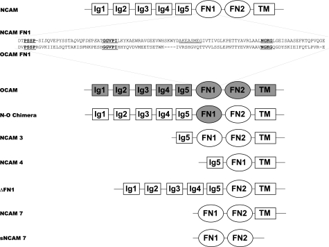
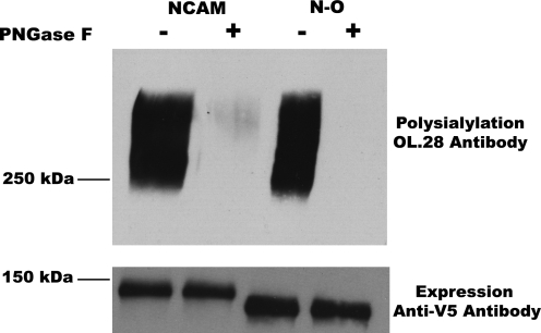
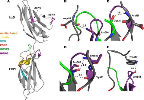
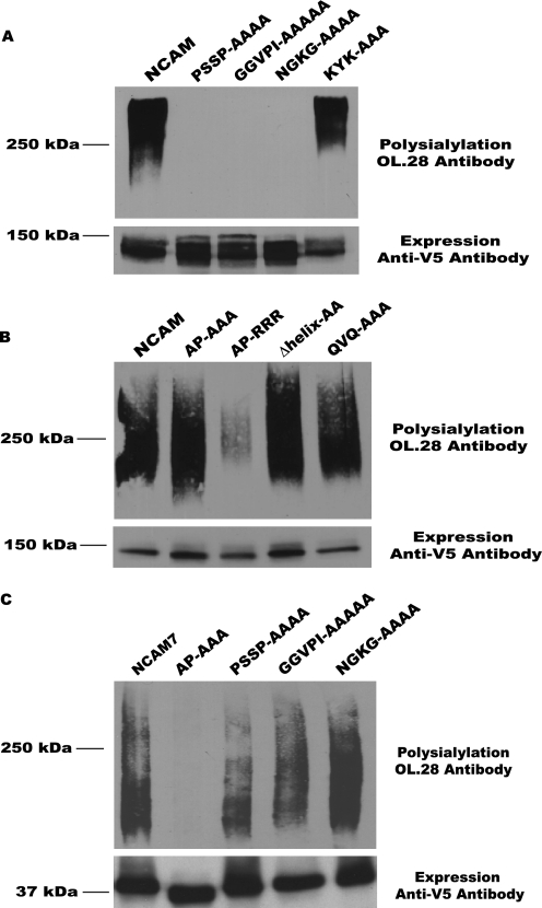
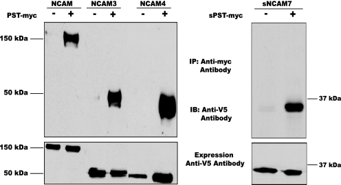
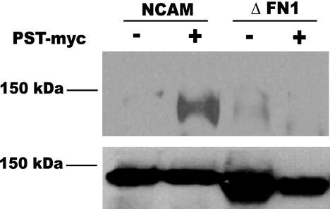
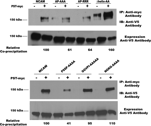
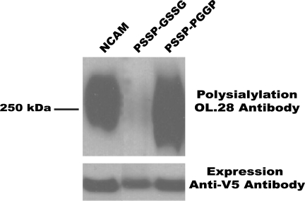
Polysialic acid is an anti-adhesive glycan that modifies a select group of mammalian proteins. The primary substrate of the polysialyltransferases (polySTs) is the neural cell adhesion molecule (NCAM). Polysialic acid negatively regulates cell adhesion, is required for proper brain development, and is expressed in specific areas of the adult brain where it promotes on-going cell migration and synaptic plasticity. The first fibronectin type III repeat (FN1) of NCAM is required for polysialylation of the N-glycans on the adjacent immunoglobulin-like domain (Ig5), and acidic residues on the surface of FN1 play a role in polyST recognition. Recent work demonstrated that the FN1 domain from the unpolysialylated olfactory cell adhesion molecule (OCAM) was able to partially replace NCAM FN1 (Foley, D. A., Swartzentruber, K. G., Thompson, M. G., Mendiratta, S. S., and Colley, K. J. (2010) J. Biol. Chem. 285, 35056-35067). Here we demonstrate that individually replacing three identical regions shared by NCAM and OCAM FN1, (500)PSSP(503) (PSSP), (526)GGVPI(530) (GGVPI), and (580)NGKG(583) (NGKG), dramatically reduces NCAM polysialylation. In addition, we show that the polyST, ST8SiaIV/PST, specifically binds NCAM and that this binding requires the FN1 domain. Replacing the FN1 PSSP sequences and the acidic patch residues decreases NCAM-polyST binding, whereas replacing the GGVPI and NGKG sequences has no effect. The location of GGVPI and NGKG in loops that flank the Ig5-FN1 linker and the proximity of PSSP to this linker suggest that GGVPI and NGKG sequences may be critical for stabilizing the Ig5-FN1 linker, whereas PSSP may play a dual role maintaining the Ig5-FN1 interface and a polyST recognition site.


Figures

FIGURE 1.
FIGURE 1.
Schematic representation of NCAM, the NCAM-OCAM chimera, and NCAM domain deletion mutants. The extracellular portion of NCAM is composed of five Ig-like domains and two fibronectin type III repeats. The NCAM isoform that is used in these studies is NCAM140, which also contains a TM and cytoplasmic tail. OCAM has the same domain structure as NCAM and a similar glycosylation site pattern but is unpolysialylated (44). Sequences common to NCAM and OCAM FN1 domains are bold and underlined in both FN1 sequences. Acidic patch residues (Asp506, Asp520, Glu521, and Glu523) and the QVQ sequence are shown in the NCAM FN1 sequence in italics, whereas the α-helix is underlined. An N-O chimera was created in which OCAM FN1 replaces the NCAM FN1 (N-O Chimera) (42). NCAM domain deletion proteins were previously constructed and analyzed (38, 40) and are used in this study. These include NCAM3 consisting of Ig5-FN1-FN2-TM-tail, NCAM4 consisting of Ig5-FN1-TM-tail, NCAM7 consisting of FN1-FN2-TM-tail, and a sNCAM7 containing only FN1-FN2. NCAM, NCAM3, and NCAM4 are predominantly polysialylated on N-linked glycans, whereas NCAM7 is polysialylated on O-linked glycans (38). The ΔFN1 NCAM mutant is not polysialylated (39).
FIGURE 2.
FIGURE 2.
The OCAM FN1 domain can replace the NCAM FN1 domain in the N-O chimera to allow the polysialylation of N-glycans. NCAM and the N-O chimera were transiently co-expressed with PST in COS-1 cells for 18 h. Proteins were immunoprecipitated using an anti-V5 epitope tag antibody, subjected to SDS-PAGE, and immunoblotted with the OL.28 antibody, as described under “Experimental Methods” (upper panel, Polysialylation, OL.28 antibody). Aliquots of cell lysates were immunoblotted with the anti-V5 antibody to determine relative protein expression levels (lower panel, Expression, Anti-V5 Antibody). To determine whether the polysialic acid was found on N-linked or O-linked glycans, immunoprecipitates were treated with peptide N-glycosidase F that selectively removes N-linked, but not O-linked, glycans (PNGase −/+).
FIGURE 3.
FIGURE 3.
Structure of NCAM Ig5-FN1 and predicted Ig5-FN1 interface interactions. A, the crystal structure of the NCAM Ig5-FN1 tandem is shown (PDB ID 3MTR (45)). The three major regions of sequence identity between NCAM FN1 and OCAM FN1, 500PSSP503 (PSSP, red), 526GGVPI530 (GGVPI, green), and 580NGKG583 (NGKG, purple) are highlighted on the FN1 sequence. NGKG and GGVPI form loops in the interface between Ig5 and FN1 domains. PSSP is in an unstructured region that connects Ig5 to FN1. Also shown are three features unique to NCAM FN1 that have been shown to be involved in the polysialylation of Ig5 N-glycans (39, 40, 42). These include the three core amino acids of an acidic surface patch, Asp520, Glu521, and Glu523 (Acidic Patch, orange), an α-helix linking strands 4 and 5 of the β sandwich (α-Helix, yellow), and the 516QVQ518 sequence (QVQ, cyan). Also shown are the two Asn residues that carry the N-glycans that are polysialylated (ASN5 and ASN6 in magenta). B, predicted hydrogen bond between the main chain carbonyl of Asp498 (Ig5-FN1 linker) and the side chain amide group of Asn580 (NGKG). C, shown is the predicted hydrogen bond between the main chain carbonyl of Gly526 (GGVPI) and the main chain amide of Asp498 (Ig5-FN1 linker). D, shown are two hydrogen bonds that stabilize the NGKG β turn; one between the side-chain carboxyl group of Asn580 and the main chain amide group of Lys582 and a second between the main-chain amide group of Asn580 and the main-chain carbonyl group of Gly583. E, shown is a possible hydrogen bond between the amine group in the indole ring of Trp417 in the Ig5 domain and the main chain carbonyl group of Gly581 of NGKG. Bond distances were predicted using the measurement function of the PyMol software.
FIGURE 4.
FIGURE 4.
Replacing sequences common to NCAM and OCAM FN1 domains dramatically reduces NCAM polysialylation. The polysialylation of NCAM and NCAM7 proteins and mutants with selected sequences replaced was analyzed by immunoblotting with the OL.28 anti-polysialic acid antibody after co-expression of NCAM or the mutant NCAM proteins with PST in COS-1 cells (upper panels, Polysialylation, OL.28 Antibody). Relative levels of protein expression were determined by immunoblotting aliquots of the cell lysates with the anti-V5 epitope tag antibody (lower panels, Expression, Anti-V5 Antibody). A, analysis of alanine replacement mutants of the sequences common to NCAM and OCAM FN1 domains (PSSP-AAAA, GGVPI-AAAAA, NGKG-AAAA, and KYK-AAA) is shown. B, analysis of previously generated mutants in the FN1 acidic patch (AP-AAA, AP-RRR) (39), α-helix (Δhelix-AA) (40), and QVQ sequence (QVQ-AAA) (42) is shown. C, analysis of NCAM7 and alanine replacement mutants of the acidic patch (AP-AAA) and sequences common to NCAM and OCAM FN1 domains is shown.
FIGURE 5.
FIGURE 5.
PST-NCAM binding does not require Ig domains or membrane association. Co-immunoprecipitation analysis of the binding of membrane-associated PST-myc and sPST-myc to NCAM and truncated NCAM proteins after co-expression in COS-1 cells is shown. Left panels, PST was immunoprecipitated from cell lysates using an anti-myc antibody, and co-immunoprecipitating proteins were detected by immunoblotting with an anti-V5 tag antibody (+PST-myc). Nonspecific binding was evaluated by performing the same analysis on lysates from cells not expressing PST-myc (−PST-myc) (upper right panel). NCAM protein expression was evaluated by immunoblotting one-tenth of the cell lysate with anti-V5 antibody (lower left panel). Right panels, sPST was immunoprecipitated (IP) from cell media using an anti-myc antibody, and co-immunoprecipitating sNCAM7 was detected by immunoblotting (IB) with an anti-V5 tag antibody (+PST-myc). Nonspecific binding was evaluated by performing the same analysis on media from cells not expressing PST-myc (−PST-myc) (upper right panels). sNCAM7 expression was evaluated by immunoblotting an aliquot of media with anti-V5 antibody (lower right panel).
FIGURE 6.
FIGURE 6.
PST-NCAM binding requires the FN1 domain of NCAM. Co-immunoprecipitation analysis of the binding of membrane associated PST-myc to NCAM and the NCAM ΔFN1 mutant is shown. Top panel, PST was immunoprecipitated from cell lysates using an anti-myc antibody, and co-immunoprecipitating proteins were detected by immunoblotting with an anti-V5 tag antibody (+PST-myc). Nonspecific binding was evaluated by performing the same analysis on lysates from cells not expressing PST-myc (−PST-myc). Lower panel, NCAM and ΔFN1 protein expression was evaluated by immunoblotting one-tenth of the cell lysate with anti-V5 antibody.
FIGURE 7.
FIGURE 7.
NCAM mutants with the acidic patch and PSSP sequences replaced demonstrate reduced binding to PST. Upper panels of each set, NCAM and its mutants with sequences of the acidic patch (AP-AAA, AP-RRR), α-helix (Δhelix-AA), PSSP (PSSP-AAAA), GGVPI (GGVPI-AAAAA), and NGKG (NGKG-AAAA) replaced were co-expressed with PST in COS-1 cells. PST was immunoprecipitated (IP) from cell lysates with an anti-myc tag antibody, and co-immunoprecipitating proteins were detected by immunoblotting (IB) with an anti-V5 epitope tag antibody (+PST-myc). Nonspecific binding was evaluated by performing the same analysis on lysates from cells not expressing PST-myc (−PST-myc). Lower panels of each set, NCAM and NCAM mutant protein expression was evaluated by immunoblotting one-tenth of the cell lysate with anti-V5 antibody. Changes in the extent of co-precipitation of mutant NCAM proteins were quantified using NIH ImageJ and are shown underneath each series of blots.
FIGURE 8.
FIGURE 8.
Pro500 and Pro503 in the PSSP sequence are critical for NCAM polysialylation. NCAM and mutants with either the two proline residues or two serine residues of the PSSP sequence replaced (PSSP-GSSG and PSSP-PGGP) were co-expressed with PST-myc in COS-1 cells. NCAM proteins were immunoprecipitated using an anti-V5 epitope tag antibody and immunoblotted with the OL.28 antibody to detect polysialylation (upper panel, Polysialylation, OL.28 Antibody). Aliquots of cell lysates were immunoblotted with the anti-V5 antibody to determine relative protein expression levels (lower panel, Expression, Anti-V5 Antibody).

Similar articles

Cited by

References

    1. Cunningham B. A., Hemperly J. J., Murray B. A., Prediger E. A., Brackenbury R., Edelman G. M. (1987) Science 236, 799–806 - PubMed
    1. Walmod P. S., Kolkova K., Berezin V., Bock E. (2004) Neurochem. Res. 29, 2015–2035 - PubMed
    1. Ditlevsen D. K., Povlsen G. K., Berezin V., Bock E. (2008) J. Neurosci. Res. 86, 727–743 - PubMed
    1. Eckhardt M., Mühlenhoff M., Bethe A., Koopman J., Frosch M., Gerardy-Schahn R. (1995) Nature 373, 715–718 - PubMed
    1. Kojima N., Yoshida Y., Tsuji S. (1995) FEBS Lett. 373, 119–122 - PubMed

Publication types

MeSH terms

Substances