. 2011 Feb 11;286(6):4566-75.
doi: 10.1074/jbc.M110.199976. Epub 2010 Dec 3.

Nuclear tau, a key player in neuronal DNA protection

Affiliations

Nuclear tau, a key player in neuronal DNA protection

Audrey Sultan et al. J Biol Chem. .

Abstract

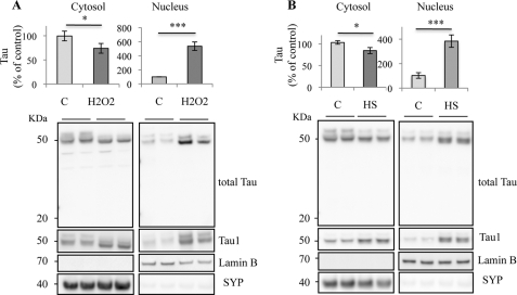
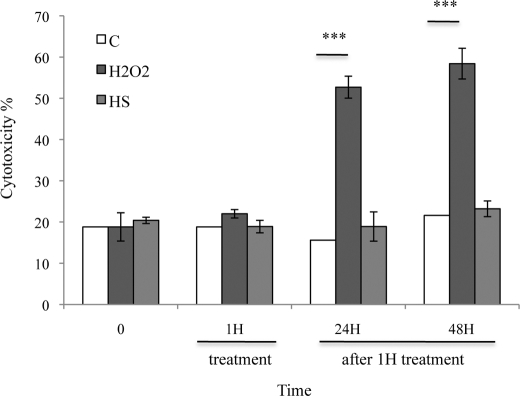
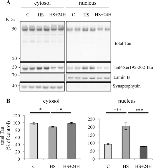
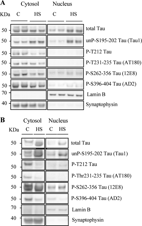
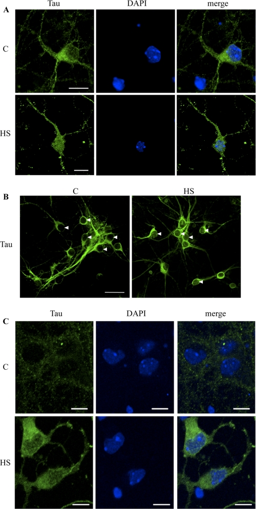
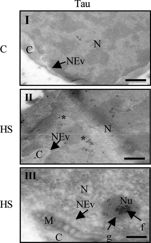
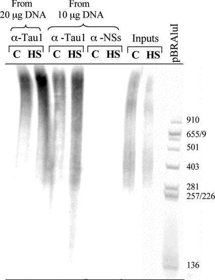
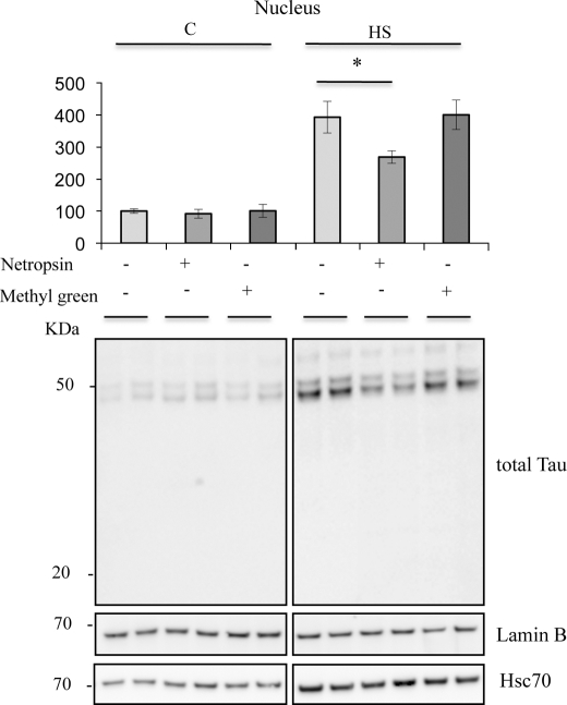
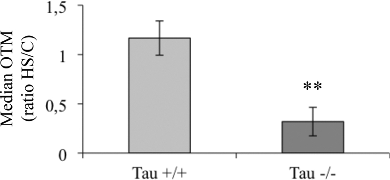
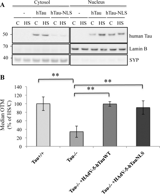
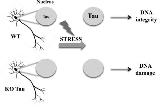
Tau, a neuronal protein involved in neurodegenerative disorders such as Alzheimer disease, which is primarily described as a microtubule-associated protein, has also been observed in the nuclei of neuronal and non-neuronal cells. However, the function of the nuclear form of Tau in neurons has not yet been elucidated. In this work, we demonstrate that acute oxidative stress and mild heat stress (HS) induce the accumulation of dephosphorylated Tau in neuronal nuclei. Using chromatin immunoprecipitation assays, we demonstrate that the capacity of endogenous Tau to interact with neuronal DNA increased following HS. Comet assays performed on both wild-type and Tau-deficient neuronal cultures showed that Tau fully protected neuronal genomic DNA against HS-induced damage. Interestingly, HS-induced DNA damage observed in Tau-deficient cells was completely rescued after the overexpression of human Tau targeted to the nucleus. These results highlight a novel role for nuclear Tau as a key player in early stress response.


Figures

FIGURE 1.
FIGURE 1.
Cellular distribution of Tau in stress-treated cortical neurons. Western blot analysis of Tau expression in primary neuronal cultures of mouse embryonic cortex using an anti-total Tau antibody recognizing the C-terminal Tau sequence and anti-nonphospho-Tau (Tau1) antibody. Cortical cultures were maintained at 37 °C (C), treated with H2O2 (A) or heated to 44 °C (HS; B) for 1 h. Cells were subjected to cellular fractionation as described under “Experimental Procedures.” Distribution of Tau was analyzed in cytosolic and nuclear fractions. H2O2 and HS increased the nuclear Tau level in cultured neurons. Densitometric analysis of Western blot using anti-total Tau antibody. Synaptophysin (SYP) and lamin B were respectively used as specific markers of the cytoplasmic and the nuclear fractions. Results are expressed as a percentage of the control. Data shown are the mean ± S.D. of four different experiments using separate cultures.
FIGURE 2.
FIGURE 2.
Effects of stress treatments on the lactate dehydrogenase level in neuronal cultures. The influence of 1 mm H2O2 and HS for 1 h or after 24- or 48-h recovery at 37 °C on lactate dehydrogenase level was measured in cultured cortical neurons. H2O2 but not HS induced lactate dehydrogenase leakage in medium 24 h after treatment.
FIGURE 3.
FIGURE 3.
Transient nuclear accumulation of Tau in heat stress-treated neurons. A, Western blotting analysis using phospho-independent (anti-C-terminal Tau) and dephospho-Tau (Tau1) antibodies. Cortical cultures were heated to 44 °C (HS) or kept at 37 °C (C) for 1 h and then maintained at 37 °C for 24 h. Total Tau and dephosphorylated Tau labeling were analyzed both in cytosolic and nuclear fractions. A loss of nuclear Tau staining was observed 24 h after HS. B, densitometric analysis of Western blot using anti-total Tau antibody. Results are expressed as percentage of the control. Data shown are the mean ± S.D. of three different experiments. ***, p = 0.0001; *, p < 0.05.
FIGURE 4.
FIGURE 4.
Heat stress modulates Tau phosphorylation in cortical neurons. A, representative Western blot analysis using anti-total Tau antibody (anti-C-terminal Tau) and phospho-dependent Tau antibodies (Tau1, PT212, AT180, 12E8, and AD2). Cortical cultures were untreated (C) or treated with HS. Tau phosphorylation at various epitopes was analyzed both in cytosolic and nuclear fractions. In the cytosol, HS induced dephosphorylation. In nuclei from HS-treated neurons, Tau accumulated in the nucleoplasm was in a dephosphorylated state. B, representative Western blot analysis using anti-total Tau antibody (anti-C-terminal Tau) and phospho-dependent antibodies (Tau1, PT212, AT180, 12E8, AD2) in brain slices from adult mouse cortex. Cortical slices were untreated or treated with HS. In adult neurons, HS induced nuclear dephosphorylation of all Tau epitopes except for Ser262–356 (12E8).
FIGURE 5.
FIGURE 5.
Confocal microscopy analysis of Tau localization in cultured neurons. A, immunofluorescence staining of total Tau (Tau3R) in cortical neuronal cultures. Tau3R antibody specifically recognizes Tau isoforms presenting three repeat domains, this being the case of the embryonic Tau isoform, independently of their phosphorylation state. Immunolabeling of Tau and DAPI nuclear counterstaining were analyzed by confocal laser scanning microscopy. HS strongly increased Tau staining in neuronal nuclei. Scale bars indicate 20 μm. B, immunofluorescence staining of total Tau (TauC17). Cells were permeabilized before ice cold methanol fixation to only visualize nuclear bound Tau. Arrowheads indicate neuronal nuclei. Nuclear fluorescence was analyzed by confocal laser scanning microscopy. C, immunofluorescence staining of nonphospho-Tau (Ser195–202) using Tau1 antibody in cortical culture of neurons. Immunolabeling of Tau1 and ToPro3 nuclear counterstaining were analyzed by confocal laser scanning microscopy. Heat shock strongly increased Tau1 staining in neuronal cytosol and nucleus. Scale bars indicate 10 μm.
FIGURE 6.
FIGURE 6.
Electron microscopy analysis of Tau localization in cultured neurons. Immunogold electron microscopy of total Tau (Tau5 antibody) in primary neuronal cultures of mouse embryo cortex. M, mitochondria; C, cytosol; N, nucleus; Nu, nucleolus; f, fibrillar area; g, granular area; NEv, nuclear envelope. Cortical cultures were untreated (C, I) or treated with HS (I, III). Asterisks indicate Tau staining. Scale bars indicate 100 nm.
FIGURE 7.
FIGURE 7.
Interaction of nuclear Tau with genomic DNA is enhanced after HS treatment. Equal amounts (20 or 10 μg) of genomic DNA from control (C) and heat-shocked (HS) primary cultured neurons were immunoprecipitated with Tau1 antibody or an antibody directed against the viral nonstructural NSs protein (anti-NSs used here as a negative control). Immunoprecipitated and input DNA (corresponding to nonimmunoprecipitated genomic DNA) as well as pBR322/AluI DNA were 5′-end 32P-radioactively labeled and submitted to nondenaturating electrophoresis. Tau interacted with DNA in a dose-dependent manner. HS increased the Tau-DNA interaction. Data shown are representative of three independent experiments using separate cultures.
FIGURE 8.
FIGURE 8.
Netropsin modulates HS-induced nuclear Tau accumulation. (Bottom), Western blot analysis of effects of netropsin on Tau, Hsc70, and lamin B expression in the nuclear fraction using, respectively, anti-total Tau (C-terminal Tau), anti-Hsc70 and anti-lamin B antibodies. (Top), densitometric analysis of Western blot using an anti-total Tau (C-terminal Tau) antibody. Results are expressed as a percentage of the control. Data shown are the mean ± S.D. of three different experiments. *, p < 0.05.
FIGURE 9.
FIGURE 9.
Heat stress selectively impairs DNA integrity in Tau-deficient neuronal cultures. The effect of HS on DNA from wild-type (Tau+/+) or Tau-deficient cells (Tau−/−) was measured by Comet assay. Results are presented as the OTM ratio from heat-shocked cells (HS) over control (C). Each OTM value is a median value from 150 to 200 cells. Data shown are the means from three different experiments using separate cultures. **, p < 0.01.
FIGURE 10.
FIGURE 10.
Heat stress-induced DNA damage induced in Tau-deficient neurons is prevented by hTau-NLS or hTau overexpression. A, Western blotting analysis using a specific antibody raised against human Tau (HT7). Tau-deficient cortical cultures were infected for 24 h with adenoviral vectors coding for human Tau (HAdV-5-hTau44Wt) or human Tau-NLS (HAdV-5-hTau44NLS), heated to 44 °C (HS) or maintained at 37 °C (C) for 1 h and subjected to subcellular fractionation. hTau-NLS was specifically expressed in the nuclei independently of temperature treatment. hTau was mostly localized in the cytosolic fraction in the control condition. Nuclear hTau level increased after HS. B, Comet assay showing the effect of human Tau and human Tau-NLS overexpression in Tau-deficient cells. Data shown are the means ± S.D. of 100 cells measured for each condition. **, p < 0.005. SYP, synaptophysin.
FIGURE 11.
FIGURE 11.
Tau-mediated DNA protection in postmitotic neurons. Shown is a scheme illustrating the nuclear Tau protection of DNA integrity in heat-stressed neurons.

References

    1. Weingarten M. D., Lockwood A. H., Hwo S. Y., Kirschner M. W. (1975) Proc. Natl. Acad. Sci. U.S.A. 72, 1858–1862 - PMC - PubMed
    1. Loomis P. A., Howard T. H., Castleberry R. P., Binder L. I. (1990) Proc. Natl. Acad. Sci. 87, 8422–8426 - PMC - PubMed
    1. Wang Y., Loomis P. A., Zinkowski R. P., Binder L. I. (1993) J. Cell Biol. 121, 257–267 - PMC - PubMed
    1. Thurston V. C., Zinkowski R. P., Binder L. I. (1996) Chromosoma 105, 20–30 - PubMed
    1. Cross D. C., Muñoz J. P., Hernández P., Maccioni R. B. (2000) J. Cell. Biochem. 78, 305–317 - PubMed

Publication types