. 2011 Feb;300(2):L295-304.
doi: 10.1152/ajplung.00134.2010. Epub 2010 Dec 3.

TGF-β regulates Nox4, MnSOD and catalase expression, and IL-6 release in airway smooth muscle cells

Affiliations

TGF-β regulates Nox4, MnSOD and catalase expression, and IL-6 release in airway smooth muscle cells

Charalambos Michaeloudes et al. Am J Physiol Lung Cell Mol Physiol. 2011 Feb.

Abstract

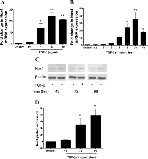
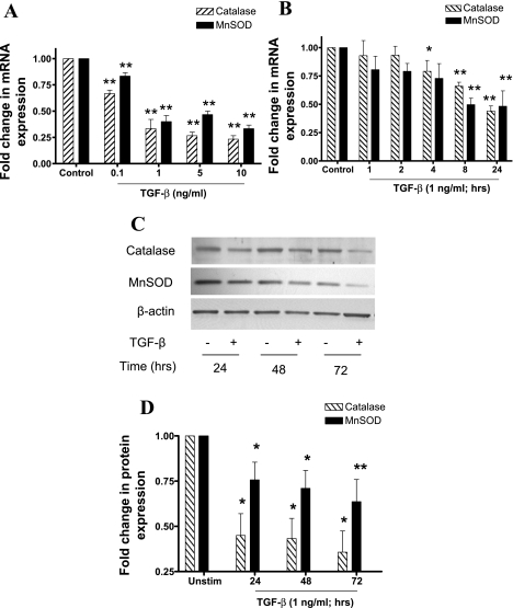
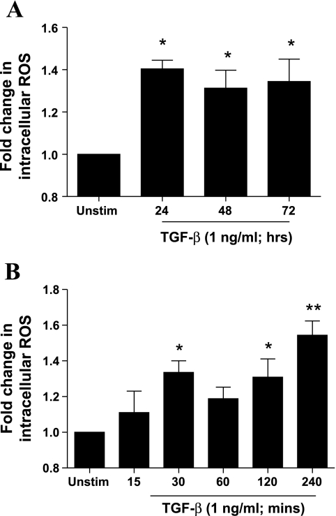
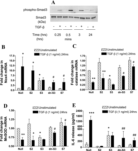
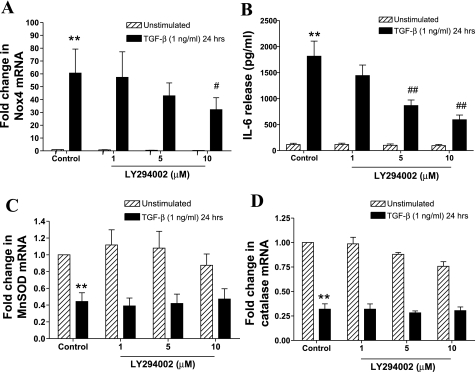
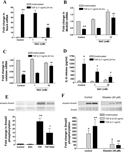
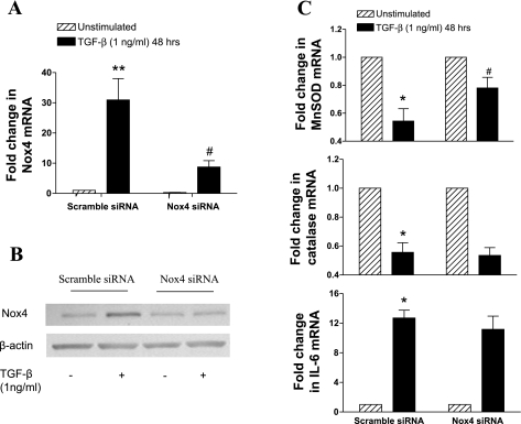
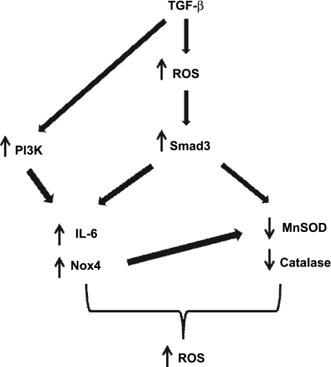
Reactive oxygen species (ROS) are generated as a result of normal cellular metabolism, mainly through the mitochondria and peroxisomes, but their release is enhanced by the activation of oxidant enzymes such as NADPH oxidases or downregulation of endogenous antioxidant enzymes such as manganese-superoxide dismutase (MnSOD) and catalase. Transforming growth factor-β (TGF-β), found to be overexpressed in airway smooth muscle (ASM) from asthmatic and chronic obstructive pulmonary disease patients, may be a pivotal regulator of abnormal ASM cell (ASMC) function in these diseases. An important effect of TGF-β on ASMC inflammatory responses is the induction of IL-6 release. TGF-β also triggers intracellular ROS release in ASMCs by upregulation of NADPH oxidase 4 (Nox4). However, the effect of TGF-β on the expression of key antioxidant enzymes and subsequently on oxidant/antioxidant balance is unknown. Moreover, the role of redox-dependent pathways in the mediation of the proinflammatory effects of TGF-β in ASMCs is unclear. In this study, we show that TGF-β induced the expression of Nox4 while at the same time inhibiting the expression of MnSOD and catalase. This change in oxidant/antioxidant enzymes was accompanied by elevated ROS levels and IL-6 release. Further studies revealed a role for Smad3 and phosphatidyl-inositol kinase-mediated pathways in the induction of oxidant/antioxidant imbalance and IL-6 release. The changes in oxidant/antioxidant enzymes and IL-6 release were reversed by the antioxidants N-acetyl-cysteine (NAC) and ebselen through inhibition of Smad3 phosphorylation, indicating redox-dependent activation of Smad3 by TGF-β. Moreover, these findings suggest a potential role for NAC in preventing TGF-β-mediated pro-oxidant and proinflammatory responses in ASMCs. Knockdown of Nox4 using small interfering RNA partially prevented the inhibition of MnSOD but had no effect on catalase and IL-6 expression. These findings provide novel insights into redox regulation of ASM function by TGF-β.


Figures

Fig. 1.
Fig. 1.
The effect of transforming growth factor (TGF)-β on NADPH oxidase 4 (Nox4) expression. A and B: abnormal airway smooth muscle cells (ASMCs) were stimulated with TGF-β (A; 0.1–10 ng/ml) for 24 h or with TGF-β (B; 1 ng/ml) for different times over a 48-h period. Data are expressed as fold change in Nox4 mRNA normalized to 18S rRNA with respect to unstimulated cells (Unstim). C and D: ASMCs were incubated with TGF-β (1 ng/ml) for different times over a 96-h period. Nox4 protein expression was determined in whole cell lysates by Western blotting (C), and band densities were measured by densitometric analysis (D). For each time point data are expressed as fold change in Nox4 protein expression normalized to β-actin expression, with respect to unstimulated control. In C the samples were loaded on the gel in a different order. For simplicity, the picture of the blot has been cut and rearranged so that the treatment times are in ascending order. Bars represent means ± SE of 3 ASMC donors (A), 8 ASMC donors (B), and 3 ASMC donors (C). *P < 0.05; **P < 0.01 compared with unstimulated control.
Fig. 2.
Fig. 2.
The effect of TGF-β on catalase and manganese-superoxide dismutase (MnSOD) expression. A and B: ASMCs were stimulated with TGF-β (A; 0.1–10 ng/ml) for 24 h or TGF-β (B; 1 ng/ml) for different times over a 24-h period. Data are expressed as fold change in mRNA expression normalized to 18S rRNA with respect to unstimulated cells. C and D: ASMCs were incubated with TGF-β (1 ng/ml) for different times over a 72-h period. MnSOD and catalase protein expression was determined in whole cell lysates by Western blotting (C), and band densities were measured by densitometric analysis (D). For each time point, data are expressed as fold change in protein expression normalized to β-actin expression, with respect to unstimulated control. Bars represent means ± SE of 3 ASMC (A), 5 ASMC donors (B), and 6 ASMC donors (C). *P < 0.05 and **P < 0.01 compared with unstimulated control.
Fig. 3.
Fig. 3.
Effect of TGF-β on intracellular reactive oxygen species (ROS) levels and IL-6 release. ASMCs were incubated with TGF-β (1 ng/ml) for different times over a 72-h period (A) or 240-min period (B). At the end of the stimulation period cells were incubated with 2′-7′-dichlorodihydrofluorescein diacetate (10 μM) and fluorescence was measured at 485/530 nm and normalized to changes in cell number as determined by methylthiazolyldiphenyl-tetrazolium bromide assay. Data shown represent relative fluorescence units normalized for cell number and are expressed as fold change with respect to unstimulated control. Bars represent means ± SE of 3 ASMC donors (A) and 5 ASMC donors (B). *P < 0.05 and **P < 0.01 compared with unstimulated control.
Fig. 4.
Fig. 4.
Role of Smad-dependent signaling in the regulation of Nox4, MnSOD, catalase expression, and IL-6 release by TGF-β. A: effect of TGF-β on Smad3 phosphorylation over time. ASMCs were treated with TGF-β (1 ng/ml) for different times over a 24-h period. Phosphorylated and total Smad3 protein expression was determined in whole cell lysates by Western blotting. The blot is representative of experiments on 4 ASMC donors. BD: ASMCs were infected with null virus (Null), wt-Smad2 (S2), wt-Smad3 (S3), wt-Smad7 (S7), or dn-Smad3 (dn-S3) adenoviral constructs for 72 h and stimulated with TGF-β (1 ng/ml) for the last 24 h of the infection period. Data are expressed as fold change in Nox4 (B), catalase (C), MnSOD (D) mRNA expression, and IL-6 release (E) with respect to null virus control. Bars represent means ± SE of 3 ASMC donors. *P < 0.05, **P < 0.01, ***P < 0.001 compared with null; #P < 0.05 and ##P < 0.01 compared with null, TGF-β-stimulated cells.
Fig. 5.
Fig. 5.
Role of phosphatidylinositol 3-kinase (PI3K)-dependent signaling in the regulation of Nox4, MnSOD and catalase mRNA expression, and IL-6 release by TGF-β. ASMCs were pretreated with LY294002 (1–10 μM) for 1 h and then incubated with TGF-β (1 ng/ml) for 24 h. Nox4 (A), MnSOD (C), and catalase (D) mRNA expression was determined by real-time PCR and expressed as fold change with respect to unstimulated control. IL-6 release (B) was determined by ELISA. Bars represent means ± SE of 4 ASMC donors **P < 0.01 compared with unstimulated control; #P < 0.05 and ##P < 0.01 compared with TGF-β-stimulated cells.
Fig. 6.
Fig. 6.
Effect of the antioxidant N-acetyl-cysteine (NAC) on the regulation of Nox4, MnSOD and catalase mRNA expression, and IL-6 release by TGF-β. AD: AMSCs were pretreated with NAC (1–10 mM) for 1 h and then incubated with TGF-β (1 ng/ml) for 24 h. Nox4 (A), MnSOD (B) and catalase (C) mRNA expression, and IL-6 release (D) were determined. E and F: ASMCs were pretreated with NAC (10 mM; E) or ebselen (40 μM; F) for 1 h and then stimulated with TGF-β (0.25–1 ng/ml) for 15 min. Phosphorylated and total Smad3 protein expression was determined in whole cell lysates by Western blotting. Data are expressed as fold change in phosphorylated-Smad3 protein expression normalized to total Smad3 expression, with respect to unstimulated control. In the experiments using ebselen, the effect of different concentrations (10, 20, and 40 μM) of the antioxidant on Smad3 phosphorylation was determined. Due to their large number the samples were loaded on 2 different gels. For simplicity only the sections of the blots showing the effect of control and 40 μM of ebselen are depicted in F. Bars represent means ± SE of 3 ASMC donors. *P < 0.05 and **P < 0.01 compared with unstimulated control; #P < 0.05 and ##P < 0.01 compared with TGF-β (1 ng/ml)-stimulated cells; $P < 0.01 compared with TGF-β (0.25 ng/ml)-stimulated cells.
Fig. 7.
Fig. 7.
Effect of Nox4 small-interfering RNA (siRNA) on the regulation of MnSOD, catalase, and IL-6 expression by TGF-β. AMSCs were transfected with Nox4 siRNA (250 nM) for 18 h, serum deprived for 6 h, and then incubated with TGF-β (1 ng/ml) for 48 h. Nox4 mRNA was determined by real-time PCR (A), and protein was determined in whole cell extracts by Western blotting (B). MnSOD, catalase, and IL-6 (C) mRNA were determined by real-time PCR. mRNA data were expressed as fold change with respect to unstimulated control. Bars represent means ± SE of 4 ASMC donors. *P < 0.05 and **P < 0.01 compared with unstimulated control; #P < 0.05 compared with TGF-β (1 ng/ml)-stimulated cells.
Fig. 8.
Fig. 8.
Schematic summary of the proposed mechanism for TGF-β-mediated regulation of oxidant/antioxidant balance and IL-6 release in ASMCs. Early release of ROS in response to TGF-β activates the Smad pathway leading to upregulation of Nox4 and IL-6 expression and downregulation of catalase and MnSOD expression. Activation of PI3K also contributes to the upregulation of Nox4 and IL-6. The upregulation of Nox4 may also contribute to the regulation of MnSOD expression but not of catalase or of IL-6. The resulting oxidant/antioxidant imbalance leads to increased levels of intracellular ROS.

References

    1. Ammit AJ, Moir LM, Oliver BG, Hughes JM, Alkhouri H, Ge Q, Burgess JK, Black JL, Roth M. Effect of IL-6 trans-signaling on the pro-remodeling phenotype of airway smooth muscle. Am J Physiol Lung Cell Mol Physiol 292: L199–L206, 2007 - PubMed
    1. Bakin AV, Stourman NV, Sekhar KR, Rinehart C, Yan X, Meredith MJ, Arteaga CL, Freeman ML. Smad3-ATF3 signaling mediates TGF-beta suppression of genes encoding Phase II detoxifying proteins. Free Radic Biol Med 38: 375–387, 2005. - PubMed
    1. Black D, Lyman S, Qian T, Lemasters JJ, Rippe RA, Nitta T, Kim JS, Behrns KE. Transforming growth factor beta mediates hepatocyte apoptosis through Smad3 generation of reactive oxygen species. Biochimie 89: 1464–1473, 2007 - PMC - PubMed
    1. Brar SS, Kennedy TP, Whorton AR, Murphy TM, Chitano P, Hoidal JR. Requirement for reactive oxygen species in serum-induced and platelet-derived growth factor-induced growth of airway smooth muscle. J Biol Chem 274: 20017–20026, 1999 - PubMed
    1. Catley MC, Sukkar MB, Chung KF, Jaffee B, Liao SM, Coyle AJ, Haddad el-B, Barnes PJ, Newton R. Validation of the anti-inflammatory properties of small-molecule IkappaB Kinase (IKK)-2 inhibitors by comparison with adenoviral-mediated delivery of dominant-negative IKK1 and IKK2 in human airways smooth muscle. Mol Pharmacol 70: 697–705, 2006 - PubMed

Publication types

MeSH terms

LinkOut - more resources