. 2011 Mar;10(3):302-12.
doi: 10.1128/EC.00201-10. Epub 2010 Dec 3.

The fungal type II myosin in Penicillium marneffei, MyoB, is essential for chitin deposition at nascent septation sites but not actin localization

Affiliations

The fungal type II myosin in Penicillium marneffei, MyoB, is essential for chitin deposition at nascent septation sites but not actin localization

David Cánovas et al. Eukaryot Cell. 2011 Mar.

Abstract

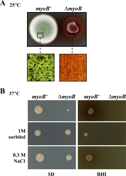
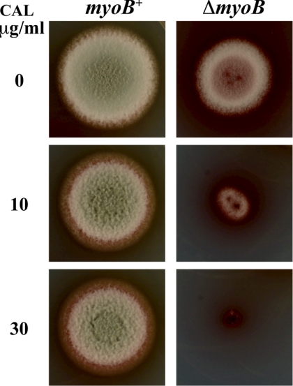
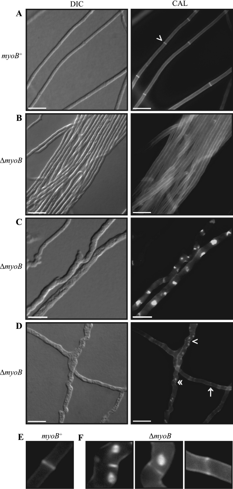
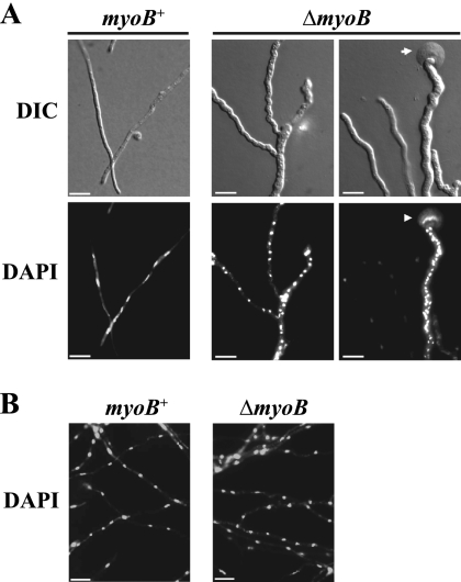
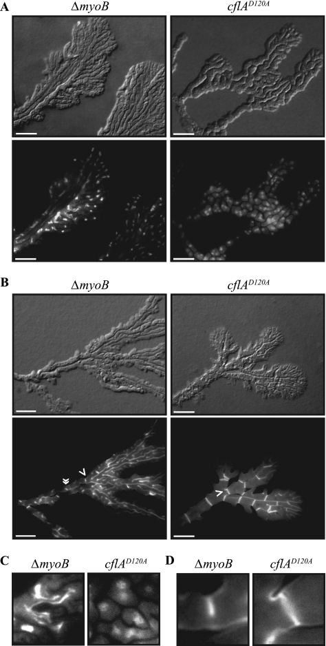
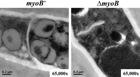
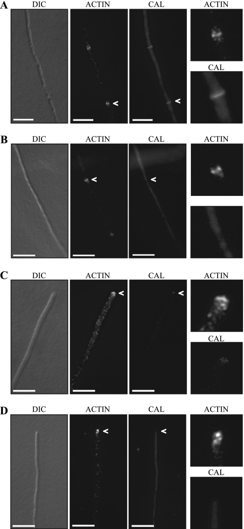
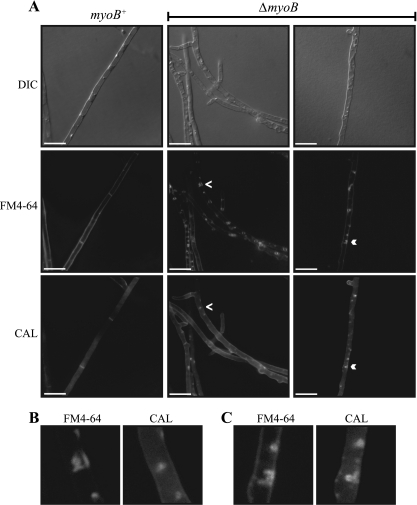
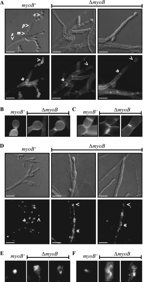
Cytokinesis is essential for proliferative growth but also plays equally important roles during morphogenesis and development. The human pathogen Penicillium marneffei is capable of dimorphic switching in response to temperature, growing in a multicellular filamentous hyphal form at 25°C and in a unicellular yeast form at 37°C. P. marneffei also undergoes asexual development at 25°C to produce multicellular differentiated conidiophores. Thus, P. marneffei exhibits cell division with and without cytokinesis and division by budding and fission, depending on the cell type. The type II myosin gene, myoB, from P. marneffei plays important roles in the morphogenesis of these cell types. Deletion of myoB leads to chitin deposition defects at sites of cell division without perturbing actin localization. In addition to aberrant hyphal cells, distinct conidiophore cell types are lacking due to malformed septa and nuclear division defects. At 37°C, deletion of myoB prevents uninucleate yeast cell formation, instead producing long filaments resembling hyphae at 25°C. The ΔmyoB cells also often lyse due to defects in cell wall biogenesis. Thus, MyoB is essential for correct morphogenesis of all cell types regardless of division mode (budding or fission) and defines differences between the different types of growth.


Figures

Fig. 1.
Fig. 1.
Deletion of the myoB gene leads to growth defects. (A) Deletion of the myoB head domain and part of the tail domain by homologous recombination leads to defects in vegetative hyphal growth at 25°C. The wild-type (myoB+) and ΔmyoB strains were grown on ANM for 8 days at 25°C. In comparison to the wild type, the ΔmyoB strain shows a growth defect (reduced colony diameter) and significantly reduced asexual development (lack of green coloration due to the absence of mature asexual spores). The lower panels show a magnified region of the surface of the colony and the lack of the mature conidiophore structures in the ΔmyoB strain. (B) Deletion of the ΔmyoB gene leads to defects in yeast morphogenesis and/or vegetative yeast growth at 37°C. The wild-type (myoB+) and ΔmyoB strains were grown on either SD or BHI medium for 6 days at 37°C with and without the addition of 1 M sorbitol or 0.3 M NaCl as an osmotic stabilizing agent. On SD medium, the ΔmyoB strain shows significantly slower growth, which is partially remediated by the addition of sorbitol or NaCl. On BHI medium, the ΔmyoB strain is completely inhibited, and this is cannot be suppressed by the addition of sorbitol or NaCl.
Fig. 2.
Fig. 2.
The ΔmyoB strain shows increased sensitivity to calcofluor. The wild-type (myoB+) and ΔmyoB strains were grown on ANM for 8 days at 25°C in the presence or absence of different concentrations of the chitin binding agent calcofluor (CAL). The ΔmyoB strain is significantly more sensitive to calcofluor than the wild type.
Fig. 3.
Fig. 3.
The ΔmyoB strain shows defects in chitin deposition. The wild-type (myoB+) (A and E) and ΔmyoB (B to D and F) strains were grown for 4 days at 25°C and stained with calcofluor (CAL) to visualize chitin deposition in cell walls and septa. (A) In the wild type, septa (arrowhead) are observed at regular intervals along hyphae, separating the cellular compartments. (B) In contrast to those of the wild type, the hyphae of the ΔmyoB strain clump together and form hyphal cables. (C) Large, abnormal deposits of chitin are observed along the hyphae of the ΔmyoB strain. (D) Aberrant septa are observed in the ΔmyoB strain. Incompletely formed septa are observed, with chitin on only one side (single arrowhead) or as two separated chitin spots on either side of the hyphae (arrow). Weakly stained complete septa are also observed (double arrowheads). (E) Magnification of the wild-type septum indicated by a single arrowhead in panel A. (F) Magnification of the ΔmyoB aberrant septa indicated in panel D. Scale bars, 20 μm.
Fig. 4.
Fig. 4.
Aberrant branching and nuclear distribution at the hyphal tips of the ΔmyoB strain. The wild-type (myoB+) and ΔmyoB strains were grown on ANM for 2 days or 4 days at 25°C and then stained with 4,6-diamidino-2-phenylindole (DAPI) to visualize nuclei. (A) Apical cells of the wild-type (myoB+) strain show highly polarized growth without apical branching and regular distribution of nuclei, whereas many apical cells from the ΔmyoB strain display irregular morphology due to aberrant polarized growth, which is also evident as apical cell branching. In addition, the tips of some apical cells lyse (arrowhead), and these cells contain a large number of DAPI-stained nuclei. (B) Subapical cells from both the myoB+ and ΔmyoB strains often show regular distribution of nuclei despite the lack of normal septation in the ΔmyoB strain. Scale bars, 20 μm.
Fig. 5.
Fig. 5.
The ΔmyoB strain shares phenotypes with the dominant negative cflA mutants at 25°C. The ΔmyoB and cflAD120A mutant strains were grown at 25°C on ANM for 7 days and stained with Hoechst 33258 (A and C) or calcofluor (B and D). (A and B) Both the ΔmyoB and cflAD120A mutant strains produce apical cells with aberrant morphology. Apical cells become multibranched and appear fused. (A) In the ΔmyoB strain, these structures contain nuclei with aberrant morphology, suggestive of nuclear division defects. In contrast, additional nuclei are observed in the cflAD120A mutant, and these appear to be normal in morphology. (B) The ΔmyoB strain has no or aberrant chitin deposition at septal sites. An incomplete septum is indicated by the single arrowhead, and an incomplete septum with a serpentine appearance is indicated by the double arrowheads. In contrast to the case for the ΔmyoB strain, the aberrant apical cells in the cflAD120A mutant have numerous septa. (C) Magnified region from panel A, showing fragmented nuclei in the ΔmyoB strain compared to nuclei of normal morphology in the cflAD120A mutant. (D) Magnified region indicated by the single arrowheads in panel B, showing the incomplete septa formed in the ΔmyoB strain compared to the formation of normal septa in the cflAD120A mutant. Scale bars, 20 μm.
Fig. 6.
Fig. 6.
The ΔmyoB strain shows incomplete septation. Transmission electron microscopy (TEM) was performed on hyphal cells of the wild-type (myoB+) and ΔmyoB strains grown on ANM for 8 days at 25°C. Comparisons of longitudinal sections show that the ΔmyoB strain produces incomplete or poorly defined septa compared to the wild-type (myoB+) strain.
Fig. 7.
Fig. 7.
myoB is not required for actin localization at nascent septation sites. The wild-type (myoB+) and ΔmyoB strains were grown on ANM for 4 days at 25°C. Chitin and actin distributions were examined by calcofluor staining (CAL) and immunocytochemistry. Magnified images are shown in the far right panels. (A) In the wild type, actin is localized as cortical actin spots along hyphae and at nascent septation sites (arrowheads). Actin localization at nascent septation sites occurs concomitantly with chitin deposition (arrowheads). (B) Actin is concentrated at nascent septation sites in the ΔmyoB strain; however, no chitin is deposited at this site. (C) In the wild type, actin is concentrated at the hyphal apex (arrowheads). (D) Actin concentrated at the hyphal apex is also observed in the ΔmyoB strain. Scale bars, 20 μm.
Fig. 8.
Fig. 8.
Distinct cellular compartments are not produced in the ΔmyoB mutant. The wild-type (myoB+) and ΔmyoB strains were grown on ANM for 3 days at 25°C and costained for calcofluor (CAL) and the membrane dye FM4-64. Single-dye controls showed no background in either channel. (A) In the wild type, the outer plasma membrane and membranes at septation sites are visible. In contrast, the membranes at possible septation sites are not visible in the ΔmyoB strain; rather, a large accumulation of circular membranes is observed along the hyphae. A proportion of these membranes colocalize with the mislocalized chitin observed in CAL staining (single and double arrowheads). (B and C) Magnification of the regions indicated by the single arrowhead (B) and double arrowhead (C) in panel A, showing colocalization of membranes and chitin in the ΔmyoB strain. Scale bars, 20 μm.
Fig. 9.
Fig. 9.
myoB is required for asexual reproduction. The wild-type (myoB+) and ΔmyoB strains were grown on ANM with 0.1% glucose for 7 days at 25°C and stained with calcofluor (CAL) (A to C) or Hoechst 33258 (D to F). (A) Wild-type conidiophores contain a stalk (s) and several metulae (m), phialides (p), and chains of conidia (c). Each cell type is defined by a chitin septal boundary. In contrast, the conidiophores produced by the ΔmyoB strain have an aberrant morphology and lack clearly defined cell types. A rudimentary conidiophore with one or two phialides and a single terminal conidium is produced. (B) Magnification of the region indicated by single arrowheads in panel A. In the wild type, each conidium is separated by a chitin-containing septum. Some conidiophores of the ΔmyoB strain completely lack the chitin separation between the phialide and the conidium, whereas others have partially formed septa. (C) Magnification of the region indicated by double arrowheads in panel A. Incomplete septa are observed in the conidiophores of the ΔmyoB strain. (D) Sterigmata cells of a wild-type conidiophore are uninucleate. Nuclei in the conidiophores of the ΔmyoB strain showed an aberrant morphology and positioning in the cell, often with a fragmented and stringy appearance indicating division defects. (E) Magnification of the region indicated by single arrowheads in panel D. The nuclei in conidia of the wild type are uniform in shape. In contrast, nuclei in the terminal conidia of the ΔmyoB strain show aberrant morphology. (F) Magnification of the region indicated by single arrowheads in panel D. The nuclei in phialides of the wild type are uniform in shape. In contrast, nuclei in the phialides of the ΔmyoB strain show aberrant morphology. Scale bars, 20 μm.

Similar articles

Cited by

References

    1. Almonacid M., et al. 2009. Spatial control of cytokinesis by Cdr2 kinase and Mid1/anillin nuclear export. Curr. Biol. 19:961–966 - PubMed
    1. Altschul S. F., Gish W., Miller W., Myers E. W., Lipman D. J. 1990. Basic local alignment search tool. J. Mol. Biol. 215:403–410 - PubMed
    1. Andrianopoulos A. 2002. Control of morphogenesis in the human fungal pathogen Penicillium marneffei. Int. J. Med. Microbiol. 292:331–347 - PubMed
    1. Berg J. S., Powell B. C., Cheney R. E. 2001. A millennial myosin census. Mol. Biol. Cell 12:780–794 - PMC - PubMed
    1. Berger B., et al. 1995. Predicting coiled coils by use of pairwise residue correlations. Proc. Natl. Acad. Sci. U. S. A. 92:8259–8263 - PMC - PubMed

Publication types

LinkOut - more resources