. 2011 Apr 1;183(7):856-64.
doi: 10.1164/rccm.201004-0654OC. Epub 2010 Dec 3.

Segmental allergen challenge alters multimeric structure and function of surfactant protein D in humans

Affiliations

Segmental allergen challenge alters multimeric structure and function of surfactant protein D in humans

Elena N Atochina-Vasserman et al. Am J Respir Crit Care Med. .

Abstract

Rationale: Surfactant protein D (SP-D), a 43-kD collectin, is synthesized and secreted by airway epithelia as a dodecamer formed by assembly of four trimeric subunits. We have previously shown that the quaternary structure of SP-D can be altered during inflammatory lung injury through its modification by S-nitrosylation, which in turn alters its functional behavior producing a proinflammatory response in effector cells.

Objectives: We hypothesized that alterations in structure and function of SP-D may occur in humans with acute allergic inflammation.

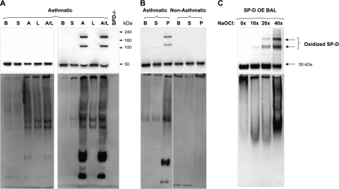
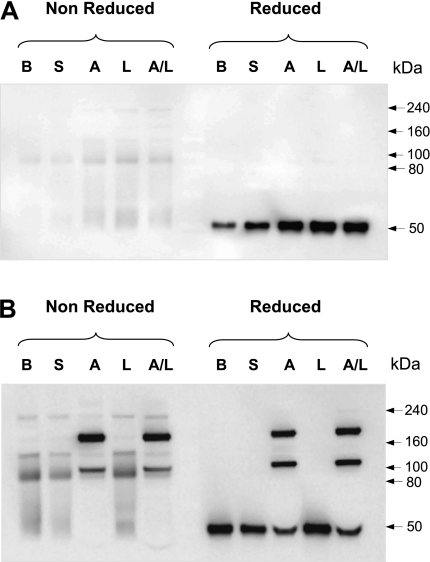
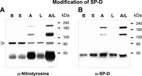
Methods: Bronchoalveolar lavage (BAL) fluid was collected from 15 nonsmoking patients with mild intermittent allergic asthma before and 24 hours after segmental provocation with saline, allergen, LPS, and mixtures of allergen and LPS. Structural modifications of SP-D were analyzed by native and sodium dodecyl sulfate gel electrophoresis.

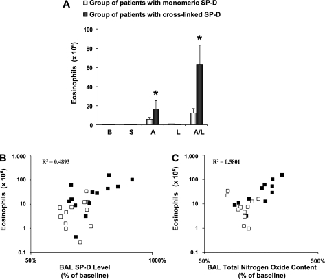
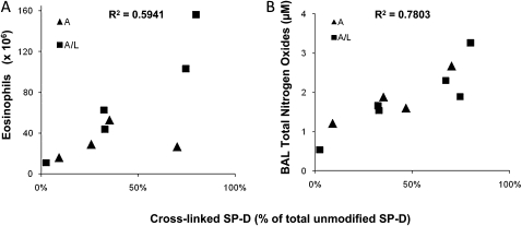
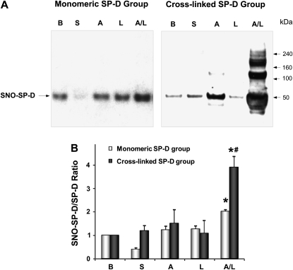
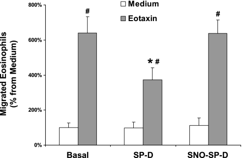
Measurements and main results: The multimeric structure of native SP-D was found to be disrupted after provocation with allergen or a mixture of allergen and LPS. Interestingly, under reducing conditions, sodium dodecyl sulfate-polyacrylamide gel electrophoresis demonstrated that 7 of 15 patients with asthma developed an abnormal cross-linked SP-D band after segmental challenge with either allergen or a mixture of allergen with LPS but not LPS alone. Importantly, patients with asthma with cross-linked SP-D demonstrated significantly higher levels of BAL eosinophils, nitrogen oxides, IL-4, IL-5, IL-13, and S-nitrosothiol-SP-D compared with patients without cross-linked SP-D.

Conclusions: We conclude that segmental allergen challenge results in changes of SP-D multimeric structure and that these modifications are associated with an altered local inflammatory response in the distal airways.


Figures

Figure 1.
Figure 1.
Challenge with allergen causes cross-linking of human surfactant protein D (SP-D) and alters its quaternary structure. (A) Blots from two subjects of the entire study group (n = 15) who underwent segmental challenges are shown. The left blots are from a representative subject who did not develop cross-linked SP-D (n = 8) and the right blots are from a representative subject who developed cross-linked SP-D (n = 7). Bronchoalveolar lavage (BAL) fluid from patients with asthma at baseline (B), and after challenge with either saline (S), house dust mite allergen (A), LPS (L), or a combination of allergen with LPS (A/L) was analyzed for total SP-D level by sodium dodecyl sulfate–polyacrylamide gel electrophoresis (SDS-PAGE) under reduced conditions (top) or by native gel electrophoresis to determine quaternary structure of SP-D (bottom). BAL from SP-D–deficient mice (SP-D−/−) was run as a negative control for nonspecific bands. Quantity of cross-linked SP-D as a percentage of total SP-D per group (A and A/L) is given in the result section and individual data are provided in Figure 4. (B) Blots from two representative subjects from a different challenge study are shown. BAL fluid from one subject with asthma (left lanes) and one healthy subject (right lanes) at baseline (B), and after challenge with saline (S) or mixed grass pollen allergen (P) were analyzed as in A. (C) Oxidative cross-linking of SP-D in vitro. BAL from mice overexpressing rat SP-D (OE BAL) was treated with various doses of NaOCl and reaction products were resolved by SDS-PAGE under reducing conditions (top) or by native gel (bottom) followed by immunoblotting with anti–SP-D antibody. Shown data are representative of five independent experiments.
Figure 2.
Figure 2.
A subset of patients displays a novel form of cross-linked surfactant protein D (SP-D). Representative blots of patients (A) who did not develop and (B) who developed abnormal cross-linked SP-D bands after segmental challenge. Bronchoalveolar lavage (BAL) from patients with asthma was denatured under reducing and nonreducing conditions and analyzed by sodium dodecyl sulfate–polyacrylamide gel electrophoresis followed by immunoblotting with anti–SP-D antibody. B = baseline; S = saline; A = allergen; L = LPS; A/L = combination of allergen with LPS. Shown data are representative of six independent experiments.
Figure 3.
Figure 3.
Patients with asthma with cross-linked surfactant protein D (SP-D) exhibit increased bronchoalveolar lavage (BAL) eosinophil numbers, SP-D, and nitrogen oxides. (A) Eosinophil cell counts after segmental challenge. The data are expressed as cell numbers × 106. Values are shown as mean ± SEM (n = 7). B = baseline; S = saline; A = allergen; L = LPS; A/L = combination of allergen with LPS. BAL eosinophils number (log converted values of the absolute number of cells, y axis) after segmental challenge with allergen and combination of allergen with LPS were correlated with (B) BAL SP-D levels (log converted values of the percent of baseline, x axis) and with (C) BAL total nitrogen oxides (percent of baseline, x axis). Mean baseline level for total nitrogen oxides was 1.41 ± 0.29 μM and 1.25 ± 0.11 μM for patients in the monomeric and cross-linked SP-D group, respectively.
Figure 4.
Figure 4.
Cross-linked surfactant protein D (SP-D) as a marker of disease severity. For the seven patients with higher molecular weight forms of SP-D, cross-linked SP-D was quantified as a percentage of total SP-D (x axis) in bronchoalveolar lavage (BAL) from segments challenged with either allergen alone (triangles, group mean 40 ± 13%) or allergen plus LPS (boxes, group mean 45 ± 11%) and was correlated with (A) BAL eosinophils number (absolute number of cells, y axis) or (B) BAL total nitrogen oxides (micromolar, y axis).
Figure 5.
Figure 5.
Cross-linked surfactant protein D (SP-D) formed by segmental challenge is nitrated in vivo. (A) Bronchoalveolar lavage (BAL) collected from patients with cross-linked SP-D was immunoprecipitated with rabbit anti–SP-D serum followed by immunoblotting for nitrotyrosine. (B) BAL from the same patient was subject to sodium dodecyl sulfate–polyacrylamide gel electrophoresis followed by immunoblotting with rabbit anti–SP-D serum. B = baseline; S = saline; A = allergen; L = LPS; A/L = combination of allergen with LPS. The arrow indicates a protein band that is not related to SP-D because it is not present in B. Shown data are representative of three independent experiments.
Figure 6.
Figure 6.
Patients with cross-linked surfactant protein D (SP-D) exhibit significantly higher S-nitrosothiol (SNO)–SP-D/SP-D ratio after segmental challenge. (A) Biotin-switch assay for SNO–SP-D content. Samples from one representative subject per group are shown. (B) Densitometric quantification of SNO–SP-D content. The data are expressed as a ratio SNO–SP-D over total SP-D (n = 7–8 in each group). *P < 0.05 from baseline level. #P < 0.05 from the corresponding challenge group. B = baseline; S = saline; A = allergen; L = LPS; A/L = combination of allergen with LPS.
Figure 7.
Figure 7.
The inhibitory effect of surfactant protein D (SP-D) on eosinophil chemotaxis can be modulated by S-nitrosylation. Eosinophils isolated from peripheral blood of allergic patients were assayed for migration toward eotaxin (positive controls), medium alone (negative control), oligomeric (native) rrSP-D, or S-nitrosylated rrSP-D combined with medium or eotaxin. The data are expressed as a percent of migrated eosinophils toward testing solutions, where 100% was calculated as the number of cells migrating toward medium only. #P < 0.05, basal migration toward eotaxin control compared with the same treatment; *P < 0.05, migration toward eotaxin control compared with rrSP-D or S-nitrosothiol–SP-D treatment. All test solutions were used in triplicate, n = 5 individual experiments.

Similar articles

Cited by

References

    1. Drickamer K. Ca2+-dependent carbohydrate-recognition domains in animal proteins. Curr Opin Struct Biol 1993;3:393–400.
    1. Voorhout WF, Veenendaal T, Kuroki Y, Ogasawara Y, Vangolde LMG, Geuze HJ. Immunocytochemical localization of surfactant protein-d (SP-D) in type-II cells, Clara cells, and alveolar macrophages of rat lung. J Histochem Cytochem 1992;40:1589–1597. - PubMed
    1. Wong CJ, Akiyama J, Allen L, Hawgood S. Localization and developmental expression of surfactant proteins D and A in the respiratory tract of the mouse. Pediatr Res 1996;39:930–937. - PubMed
    1. Wright JR. Immunoregulatory functions of surfactant proteins. Nat Rev Immunol 2005;5:58–68. - PubMed
    1. Crouch E, Persson A, Chang D, Heuser J. Molecular-structure of pulmonary surfactant protein-D (SP-D). J Biol Chem 1994;269:17311–17319. - PubMed

Publication types

MeSH terms